This video demonstrates the cell-free synthesis of nanodisc-stabilized membrane protein antigens using a dual-compartment system. The device maintains optimal conditions for efficient protein synthesis, proper folding, and stable integration into nanodiscs.
A subscription to JoVE is required to view this content. Sign in or start your free trial.
Method Article
Synthesis of Nanodisc-Stabilized Membrane Protein Antigens Using a Cell-Free System
596 views
⸱
September 26th, 2025
In This Article
Abstract
Protocol
Cell-free production of Major outer membrane protein-telodendrimer nanolipoprotein particles (MOMP-tNLPs) for subunit vaccine formulations
- Prepare MOMP-tNLPs using cell-free methods.
- Two hours before setting up the cell-free reaction, open the prokaryotic cell-free protein expression kit and thaw one of the reconstitution buffers. Once thawed, add one tablet of ethylenediaminetetraacetic acid (EDTA)-free protease inhibitor cocktail and let it dissolve fully.
Follow this protocol using a kit designed to run 5 x 1 mL reactions.
NOTE: A typical scale-up production is 3 x 1 mL.
- For each 1 mL reaction, add 525 µL of reconstitution buffer to the E. coli lysate bottle and gently roll to dissolve. Add 250 µL of reconstitution buffer to the bottle containing reaction additives (e.g., Adenosine triphosphate, Guanosine triphosphate) and gently roll to dissolve.
- Add 8.1 mL of reconstitution buffer to the reaction feed bottle, recap with a rubber stopper (take care not to touch the inside of the rubber stopper), and invert/roll gently to dissolve.
- Add 3 mL of reconstitution buffer to the amino acid mixture bottle, recap with a rubber stopper, and invert/roll gently to dissolve.
NOTE: Take care not to touch the inside of the rubber stopper as this can lead to contamination. - Add 1.8 mL of reconstitution buffer to the methionine bottle, roll gently to dissolve, and then store on ice until use.
- Prepare the reaction solution.
- To the E. coli lysate bottle, add 225 μL of reconstituted Reaction Mix, 270 μL of reconstituted amino acid mix without methionine, and 30 μL of reconstituted methionine. Additionally, add 400 μL of the 1,2-dimyristoyl-sn-glycero-3-phosphocholine (DMPC)/telodendrimer mixture, 15 μg of MOMP plasmid, and 0.6 μg of Δ49ApolipoproteinA1 plasmid. Roll/gently shake to mix.
NOTE: Ensure that both plasmids are constructed from the same plasmid backbone. Do not vortex. - Take 20 μL of the total solution and set it aside in a 1.5 mL tube for the Green fluorescent protein (GFP)-expressing control reaction (see below).
- To the E. coli lysate bottle, add 225 μL of reconstituted Reaction Mix, 270 μL of reconstituted amino acid mix without methionine, and 30 μL of reconstituted methionine. Additionally, add 400 μL of the 1,2-dimyristoyl-sn-glycero-3-phosphocholine (DMPC)/telodendrimer mixture, 15 μg of MOMP plasmid, and 0.6 μg of Δ49ApolipoproteinA1 plasmid. Roll/gently shake to mix.
- Prepare the feed solution. To the feed mix bottle, add 2.65 mL of reconstituted amino acid mix without methionine and 300 μL of reconstituted methionine. Roll/gently shake to dissolve.
NOTE: At this time, the unused reconstitution buffer and Methionine can be returned to the freezer for storage.
- Transfer 1 mL of the reaction solution to the inner reaction chamber provided in the cell-free reaction kit and seal when filled. Transfer 10 mL of the feed solution to the outer chamber of the reaction vessel and seal.
NOTE: Do not overfill the chambers! The presence of air bubbles at the top of both the inner reaction chamber and the inner feed chamber will adversely affect the reaction. Any remaining reaction solution can be placed in a 1.5 mL tube and allowed to mix alongside the main vessel.
- Add 0.5 μL of the GFP control plasmid (0.5 mg/mL) to the previously aliquoted 20 µL reaction mixture.
NOTE: Many kits are supplied with a control plasmid for quality-control purposes. Most GFP expressing plasmid with a T7 promoter and E.coli ribosome binding site (RBS) can also be used as the control plasmid.
- Place the reaction in a shaker at 300 rpm, 30 °C for up to 18 h. To verify that the reaction was successful, use a UV light source to check for fluorescence due to the synthesis of the control GFP (Figure 1) after as little as 15 min of incubation.
NOTE: These conditions, particularly temperature, may need to be optimized for the expression of other membrane proteins.
Access restricted. Please log in or start a trial to view this content.
Results
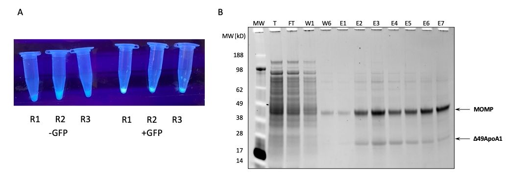
Figure 1: Expression and purification of MOMP-tNLP. (A) Image of tubes cont...
Access restricted. Please log in or start a trial to view this content.
Materials
| Name | Company | Catalog Number | Comments |
|---|---|---|---|
| 1,2-dimyristoyl-sn-glycero-3-phosphocholine (DMPC) as powder | Avanti Polar Lipids | 850345 | |
| 1.5 mL endotoxin-free centrifuge tubes | Eppendorf | 2600028 | |
| C24 Incubator shaker | New Brunswick Scientific | ||
| Cell-Free Expression System: RTS 500 ProteoMaster E. coli HY Kit | BiotechRabbit | BR1400201 | |
| cOmplete, EDTA-free Protease Inhibitor Cocktail | Roche Molecular Diagnostics | 4693132001 | |
| Orbital Shaker | |||
| PEG5K-CA8 Telodendrimer (custom synthesis product) | |||
| pIVEX2.4d vector | Roche Molecular Diagnostics | ||
| Plasmid Maxi Kit | Qiagen | 12162 | |
| UV light source |
