The video demonstrates the isolation of plastoglobules from cyanobacteria. It begins with a culture of cyanobacteria and proceeds through cell disruption using mechanical force. The release of photosynthetic pigments indicates successful lysis. A sucrose step gradient is then used to separate cellular components by density, allowing low-density plastoglobules to form a distinct band at the top. This fraction is collected and preserved for further analysis.
A subscription to JoVE is required to view this content. Sign in or start your free trial.
Method Article
Extraction and Purification of Plastoglobules from Photosynthetic Bacteria
569 views
⸱
October 30th, 2025
In This Article
Abstract
Protocol
1. Crude plastoglobule isolation
- Crude plastoglobule extraction from cyanobacteria
- Grow a 50 mL culture of Synechocystis sp. PCC 6803 to the stationary phase (about 7-10 days) and adjust the cell density to an Optical Density at 750 nm (OD750) of 2.0 using a spectrophotometer. For culture conditions, use BG-11 media and grow in an incubator at a light intensity of 150 µmol photons/m2/s, 2% CO2, 32 °C, and with continuous shaking at 150 revolutions per minute (rpm).
- To remove the polysaccharides, wash the cells by centrifuging the 50 mL culture at 6,000 × g for 45 min at 4 °C and subsequently removing the supernatant. Continue by washing the cells twice in 50 mL of buffer A (Table 1). Remove an aliquot of the cell homogenate and set it aside prior to centrifugation to be stored as a representative total cell sample
- Resuspend the washed pellet in 25 mL of Buffer A and break the cells using a French pressure cell at 1,100 pounds per square inch (psi), repeating the process 3× (put the sample on ice between each cycle to avoid protein denaturation) until the lysed colour changes from green to red-blue-green under white light. Use a pre-cooled cell and perform this step in the cold room.
- Distribute the resulting homogenate between eight 3 mL ultracentrifuge tubes filled to a maximum of 2.5 mL in each tube and then carefully overlay with 400 µL of medium R, producing a step gradient.
- Carefully balance the tubes by adding additional medium R, as necessary, and centrifuge for 30 min at 150,000 × g at 4 °C. The thylakoid and other heavier organelles (including any polyhydroxyalkanoate bodies) will pellet, while plastoglobules will be readily seen as a yellow, oily pad on or near the top of the sucrose gradient (Figure 1C).
- Harvest the resulting floating pad of crude plastoglobules with a syringe and 22G needle and deposit into a 2 mL tube. Scrape plastoglobules off the side of the ultracentrifuge tube wall with the needle tip if necessary.
- Continue to step 2.2. Alternatively, store crude plastoglobules at -80 °C and purify later.
Table 1: Buffer recipes for plastoglobule isolation from plant leaf tissue or cyanobacteria.
| Buffer compositions a | |
| Grinding Buffer | 50 mM HEPES-KOH (pH 8.0) |
| 5 mM Magnesium chloride (MgCl2) | |
| 100 mM sorbitol | |
| 5 mM ascorbic acid b | |
| 5 mM reduced cysteine b | |
| 0.05 % (w/v) Bovine Serum Albumin (BSA) b | |
| Medium R | 50 mM HEPES-KOH (pH 8.0) |
| 5 mM MgCl2 | |
| Medium R 0.2 | 50 mM HEPES-KOH (pH 8.0) |
| 5 mM MgCl2 | |
| 0.2 M sucrose | |
| Medium R 0.7 | 50 mM HEPES-KOH (pH 8.0) |
| 5 mM MgCl2 | |
| 0.7 M sucrose | |
| Buffer A | 25 mM HEPES-KOH (pH 7.8) |
| 250 mM sucrose | |
| a. Final concentrations of each buffer component are provided. | |
| b. Must be added fresh on the day of the isolation. While buffers can be prepared the day before the isolation, certain ingredients must be added fresh on the day of the isolation, as well as any phosphatase and protease inhibitors. | |
2. Harvesting pure plastoglobules
- Cyanobacteria processing
- Produce a sucrose gradient in four 2.5 mL ultracentrifuge tubes, layered first with 500 µL of the crude plastoglobules from step 1.3 mixed with 750 µL of medium R 0.7, then overlay with 750 µL of medium R 0.2.
- Centrifuge the sucrose gradient at 150,000 × g for 90 min at 4 °C. Collect the pure plastoglobules (Figure 1C) with a syringe and 22 G needle from the top phase of the sucrose gradient and transfer to a 1.5 mL tube.
- Aliquot the pure plastoglobules and flash-freeze in liquid nitrogen. Store directly at −80 °C or lyophilise to a dry powder.
Access restricted. Please log in or start a trial to view this content.
Results
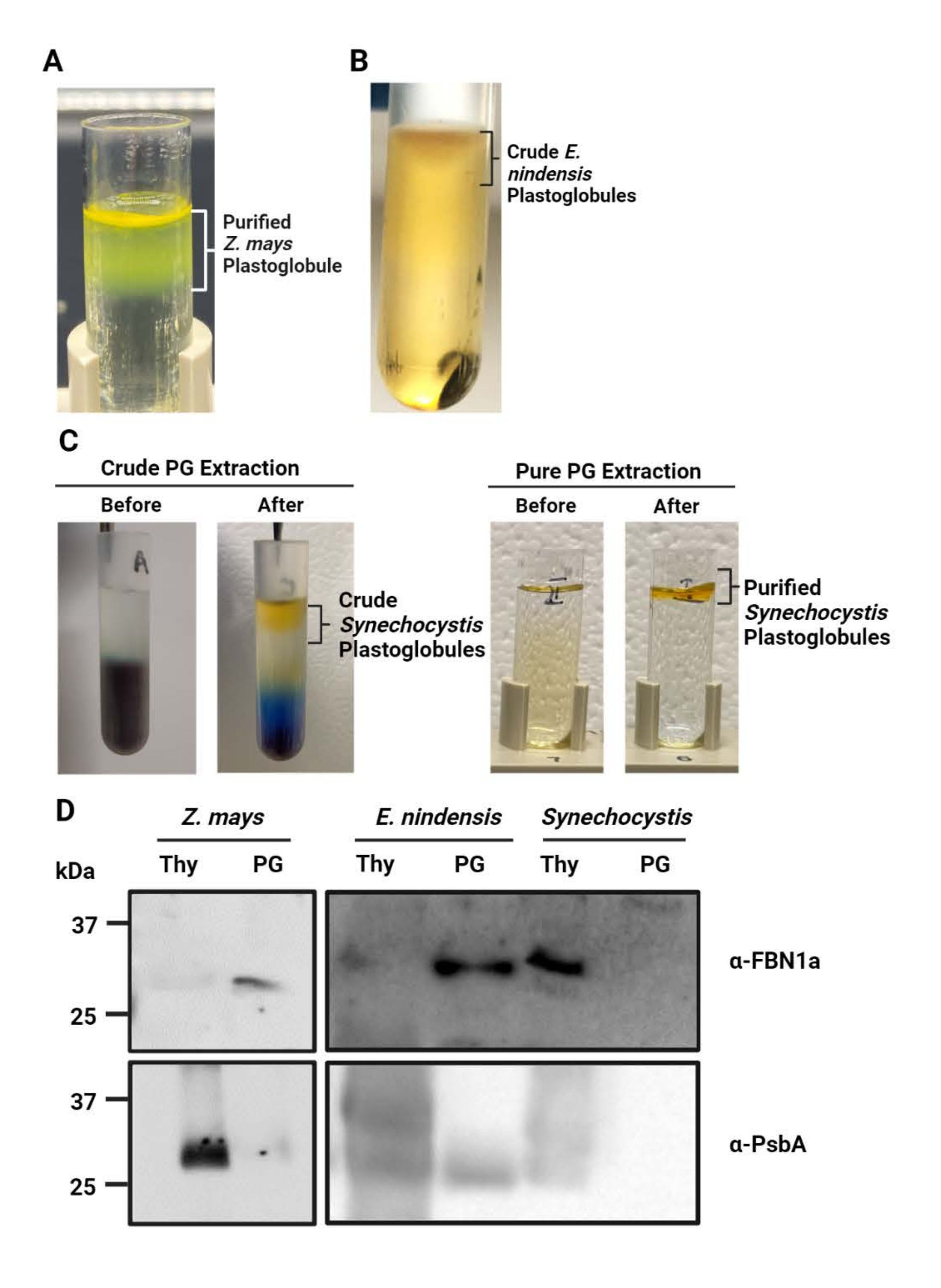
Figure 1: Isolation and immunoblotting of plastoglobules and thylakoids. (<...
Access restricted. Please log in or start a trial to view this content.
Materials
| Name | Company | Catalog Number | Comments |
|---|---|---|---|
| Ascorbate | BDH | BDH9242 | |
| Bovine Serum Albumin | Proliant Biological | 68700 | |
| French Pressure cell (model FA-079) | SLM/Aminco | N/A | |
| HEPES | Sigma Aldrich | H3375 | |
| Magnesium Chloride | Sigma Aldrich | M8266 | |
| Multitron shaking incubator | Infors HT | N/A | |
| Potassium Hydroxide | Fisher Chemicals | M16050 | |
| Reduced Cysteine | MP Biochemicals | 101444 | |
| Sorbitol | Sigma Aldrich | S3889 | |
| Sucrose | Sigma Aldrich | S9378 | |
| Synechocystis sp. PCC 6803 | N/A | N/A | |
| Optima MAX-TL Ultracentrifuge | Beckman Coulter | A95761 |
