This video demonstrates the use of a microfluidic droplet-based platform for real-time monitoring of bacterial growth. By generating uniform droplets containing bacterial suspensions and guiding them through a detection pathway, the system enables continuous optical measurement of cell density to construct a growth curve.
A subscription to JoVE is required to view this content. Sign in or start your free trial.
Method Article
Real-Time Monitoring of Bacterial Growth Using a Microfluidic Microdroplet Culture System
766 views
⸱
October 30th, 2025
In This Article
Abstract
Protocol
1. Preparations
- Connect the syringe needle (inner diameter is 0.41 mm and outer diameter is 0.71 mm), quick connector A, and reagent bottle (Figure 1C), and autoclave them at 121 °C for 15 min.
NOTE: Unscrew the cap of the reagent bottle slightly during sterilization. A few more reagent bottles can be prepared each time for use. - Use a 0.22 µm polyvinylidene fluoride (PVDF) filter to filter the microdroplet culture system (MMC) oil. Put the microfluidic chip (Figure 1B) and MMC oil into the clean bench in advance and sterilize them by ultraviolet irradiation for 30 min before use.
NOTE: For the details of Quick connector A, reagent bottle, MMC oil and microfluidic chip refer to the Table of Materials. - Install the microfluidic chip
- Open the door of the operation chamber (Figure 1A) and lift the optical fiber probe.
- Align the electric field holes with the electric field needles and gently place the chip on the chip pedestal. Then insert the two positioning columns into the positioning holes and put down the optical fiber probe (Figure 1D).
- Connect the quick connector A on the chip to the corresponding port of the MMC according to the position number (C5-O5, C4-O4, C6-O6, C2-O2, CF-OF, C1-O1, C3-O3). Then close the door of the operation chamber.
- Replenish the MMC oil (to about 80 mL) in the oil bottle and empty the waste liquid in the waste bottle before use.
NOTE: The waste liquid is usually organic waste. Please refer to regional law and regulation upon disposal, subject to change based on experimental setup.
2. Growth curve measurement in MMC
- Preparation for the initial bacterial solution
- Follow the related standard regulations to prepare Luria-Bertani (LB) medium and autoclave at 121°C for 15 min.
NOTE: Components of LB medium: NaCl (10 g/L), yeast extract (5 g/L) and tryptone (10 g/L). - Take out the Escherichia coli (E. coli) MG1655 strain from glycerol stock and cultivate it in a 50 mL shake flask with 10 mL of LB medium in a shaking incubator (200 rpm) at 37 °C for 5-8 h.
NOTE: The cultivation time depends on the specific strains. It is optimal to cultivate the strain in the logarithmic period/phase. - Dilute the cultured E. coli MG1655 solution with fresh medium to an optical density at 600 nanometers (OD600) of 0.05-0.1 to obtain an initial bacterial solution (prepare about 10 mL).
- Follow the related standard regulations to prepare Luria-Bertani (LB) medium and autoclave at 121°C for 15 min.
- Click on Initialization to initialize the MMC. After the initialization interface appears, set the cultivation temperature as 37 °C and the photoelectric signal value as 0.6 (Figure 2A). Initialization will take about 20 minutes.
- Turn on the ultraviolet (UV) lamp (wavelength 254 nm) during initialization.
- Inject the initial bacteria solution and MMC oil into the reagent bottle.
- Take out a sterilized reagent bottle on the clean bench and tighten the cap.
- Use a 10 mL sterile syringe to inject 3-5 mL of MMC oil from the syringe needle of the side tube. Tilt and rotate the reagent bottle slowly to make the oil fully infiltrate the inner wall.
- Inject about 5 mL of initial bacteria solution, and then fill the reagent bottle by injecting 5-7 mL of the oil again.
- Pull out the independent quick connector A, and insert the quick connector A of the reagent bottle into its quick connector B to complete the sample injection operation (Figure 3A).
- Wait for the initialization to end and then turn off the UV lamp (wavelength 254 nm).
- Open the door of the operation chamber, and put the reagent bottle into the metal bath.
- Pull out the C2 connector of the chip and the quick connector A of the reagent bottle. Connect the side tube connector of the reagent bottle to the C2 connector and the top tube connector to the O2 connector. Then close the door of the operation chamber.
- Click on Growth Curve to choose the function of growth curve measurement (Figure 2A). In the parameter setting interface, input the Number as 15, turn on the OD detection switch, and set the Wavelength as 600 nm. Click on Start to start droplet generation. It will take about 10 minutes.NOTE: Here, Number refers to the number of droplets to be generated. Wavelength refers to the wavelength of the OD to be detected. Set the Number (maximum 200) and Wavelength (350-800 nm) according to the experiment requirements.
- When a pop-up window appears on the main interface prompting "Remove the reagent bottle between C2 and O2, then please click the OK button after completion", open the door of the operation chamber to take out the reagent bottle and connect the C2 and O2 connectors.
- Close the door, and click the OK button in the pop-up window to automatically cultivate the droplets and detect the OD values.
NOTE: The MMC detects the OD value when the droplet passes the optical fiber probe. Therefore, the detection period depends on the number of droplets generated. - When the growth curve reaches the stationary phase, click the Data Export button to export the OD data. Select the data save path and export the OD value recorded during the cultivation period in the .csv format, which can be opened by appropriate software (e.g., Microsoft Excel). Then use a mapping software (e.g., EXCEL and Origin 9.0) to plot the growth curve.
NOTE: During the cultivation process, it is feasible to click on the Data Export at any time to export the OD data of all the current droplets.
Access restricted. Please log in or start a trial to view this content.
Results
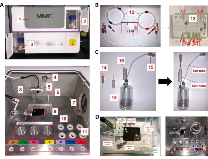
Figure 1: Structure and essential tools of MMC. (A) External and operational chamber of MMC. (B) The microfluidic chip of...
Access restricted. Please log in or start a trial to view this content.
Materials
| Name | Company | Catalog Number | Comments |
|---|---|---|---|
| 0.22 μm PVDF filter membrane | Merck Millipore Ltd. | SLGPR33RB | Sterilize the MMC oil |
| 4 °C refrigerator | Haier | BCD-289BSW | For reagent storage |
| Agar | Becton, Dickinson and Company | 214010 | For solid plate preparation |
| Clean bench | Beijing Donglian Har Instrument Manufacture Co., Ltd. | DL-CJ-INDII | For aseptic operation and UV sterilization |
| Computer | Lenovo | E450 | Software installation and MMC control |
| Constant temperature incubator | Shanghai qixin scientific instrument co., LTD | LRH 250 | For the microbial cultivation using solid medium |
| Electronic balance | OHAUS | AR 3130 | For reagent weighing |
| EP tube | Thermo Fisher | 1.5 mL | For droplet collection |
| Freezing tube | Thermo Fisher | 2.0 mL | For strain preservation |
| Glycerol | GENERAL-REAGENT | G66258A | For strain preservation |
| High-pressure steam sterilization pot | SANYO Electric | MLS3020 | For autoclaved sterilization |
| Microbial microdroplet culture system (MMC) | Luoyang TMAXTREE Biotechnology Co., Ltd. | MMC-I | Performing growth curve determination and adaptive evolution. Please refer to http://www.tmaxtree.com/en/index.php?v=news&id=110 |
| Microfluidic chip | Luoyang TMAXTREE Biotechnology Co., Ltd. | MMC-ALE-OD | For various droplet operations. Please refer to http://www.tmaxtree.com/en/ |
| MMC oil | Luoyang TMAXTREE Biotechnology Co., Ltd. | MMC-M/S-OD | The oil phase for droplet microfluidics. Please refer to http://www.tmaxtree.com/en/ |
| NaCl | GENERAL-REAGENT | G81793J | Component of the LB medium |
| Petri dish | Corning Incorporated | 90 mm | For the preparation of solid medium |
| Pipette | eppendorf | 2.5 μL, 10 μL, 100μL, 1000μL | For liquid handling |
| Quick connector A | Luoyang TMAXTREE Biotechnology Co., Ltd. | — | For the connection of each joint. Please refer to http://www.tmaxtree.com/en/ |
| Reagent bottle | Luoyang TMAXTREE Biotechnology Co., Ltd. | MMC-PCB | Sampling and storage of bacteria solution and reagents. Please refer to http://www.tmaxtree.com/en/ |
| Shake flask | Union-Biotech | 50 mL | For microbial cultivation |
| Shaking incubator | Shanghai Sukun Industrial Co., Ltd. | SKY-210 2B | For the microbial cultivation in shake flask |
| Syringe | JIANGSU ZHIYU MEDICAL INSTRUCTMENT CO., LTD | 10 mL | Draw liquid and inject it into the reagent bottle |
| Syringe needle | OUBEL Hardware Store | 22G | Inner diameter is 0.41 mm and outer diameter is 0.71 mm. |
| Tryptone | Oxoid Ltd. | LP0042 | Component of the LB medium |
| Ultra low temperature refrigerator | SANYO Ultra-low | MDF-U4086S | For strain preservation (-80 °C) |
| UV–Vis spectrophotometer | General Electric Company | Ultrospec 3100 pro | For the measurement of OD values |
| Yeast extract | Oxoid Ltd. | LP0021 | Component of the LB medium |
