The video demonstrates the delivery of fluorescent biomolecules into electrocompetent microbial cells using electroporation. The process begins with the introduction of fluorophore-tagged DNA or proteins into a chilled cell suspension. A high-voltage pulse is then applied to create temporary membrane pores, enabling biomolecule entry into the cytoplasm. Following recovery, the cells undergo washing and filtration to remove non-internalized or surface-bound molecules. Finally, the labeled cells are mounted for fluorescence imaging to visualize intracellular components.
A subscription to JoVE is required to view this content. Sign in or start your free trial.
Method Article
Electroporation-Based Delivery of Fluorescent Biomolecules into Bacterial Cells
524 views
⸱
November 28th, 2025
In This Article
Abstract
Protocol
1. Electroporation
- Incubation
- Add up to 5 µl of labeled molecules stored in a low-salt buffer (< 50 mM salt) to a single aliquot of competent cells (20 µl bacteria or 50 µl yeast) and incubate for 10 min on ice.
NOTE: The concentration of fluorescently labeled molecules in the stock solutions and thus the amount of labeled molecules added to the cell prior to electroporation are directly correlated with loading efficiency (Figure 1 and Discussion). As some proteins are less compatible with low salt conditions, the salt concentration in the storage buffer might be increased, but the volume of labeled molecules added to the cells prior to electroporation then needs to be decreased. - Transfer the mixture of cells and labeled biomolecules into a pre-chilled electroporation cuvette (0.1 and 0.2 cm spacing for bacteria and yeast, respectively). Gently tap the cuvette on the bench to remove any potential bubbles from the solution.
- Place the cuvette into the electroporator and apply a high-voltage electric pulse to the solution (0.9 to 1.8 kV/cm, see Discussion for more details on choosing voltage). Such a pulse forms transient pores in the cell membranes, allowing labeled biomolecules to diffuse into the cells.
- Check that the time constant displayed on the electroporator is between 4 to 6 ms. Lower time constants are often due to too high salt concentration and/or the presence of bubbles in the cuvette, and will lead to very low or no loading of the cells.
- Add up to 5 µl of labeled molecules stored in a low-salt buffer (< 50 mM salt) to a single aliquot of competent cells (20 µl bacteria or 50 µl yeast) and incubate for 10 min on ice.
- Recovery
- Immediately after electroporation, add 500 μl of rich medium such as Super Optimal Broth with Catabolite repression (SOC), EZ Rich Defined Medium, Yeast extract Peptone Dextrose (YPD), or any rich medium to the cells.
- Incubate the sample at 37 °C for bacteria and 29 °C for yeast for 2 to 10 min. For viability measurements, where the user wants to evaluate the percentage of cells growing and dividing after electroporation, use a longer recovery time (up to 1 hour) as we observe such lag times before the first cell division.
- Washing steps
- Wash the cells to remove any non-internalized biomolecules by spinning down the cells for 1 min at 3300 x g and 4 °C. Discard the supernatant and resuspend the cells in 500 μl Phosphate-Buffered Saline (PBS).
NOTE: For each sample, prepare a negative control of cells incubated with the same amount of labeled biomolecules but not electroporated and washed exactly the same way as the main sample. - Repeat the previous steps 3 times.
- In the case of protein internalization, optimize the washing procedure depending on the properties and behavior of the labeled protein of interest. The following steps are examples of possible optimizations:
- Perform the first three washing cycles using PBS containing 100 mM NaCl and 0.005% Triton X-100 to remove non-internalized proteins that might stick to the outer cell membrane.
- Filter the electroporated cells with a 0.22 µm pore diameter filter fitted inside a 1.5 ml microcentrifuge tube by pipetting the electroporated cells into the filter. Spin down for 3 min at 800 x g and 4 °C. Discard the flow-through. Add 500 µl of new PBS over the cells and spin them once again as before, and repeat these steps once.
- Add a small amount of protease K (10 ng in 500 μl PBS) during the first washing cycle to allow the digestion of any non-internalized protein.
- Perform the first three washing cycles using PBS containing 100 mM NaCl and 0.005% Triton X-100 to remove non-internalized proteins that might stick to the outer cell membrane.
- Spin down the cells for 1 minute at 3300 x g and 4 °C. Discard the supernatant and resuspend the cells in 150 μl of PBS.
- Spread the loaded cell solution on the agarose pad by removing the upper coverslip and spreading 10 µl of the cell suspension droplet by droplet. Replace an unused clean, burned coverslip (No 1.5 thickness, matching the microscope objective specification) on the top of the pad and press very gently on the slide.
- Protect the electroporated cells from light by storing the pads in an opaque box while imaging different samples.
- Wash the cells to remove any non-internalized biomolecules by spinning down the cells for 1 min at 3300 x g and 4 °C. Discard the supernatant and resuspend the cells in 500 μl Phosphate-Buffered Saline (PBS).
Access restricted. Please log in or start a trial to view this content.
Results
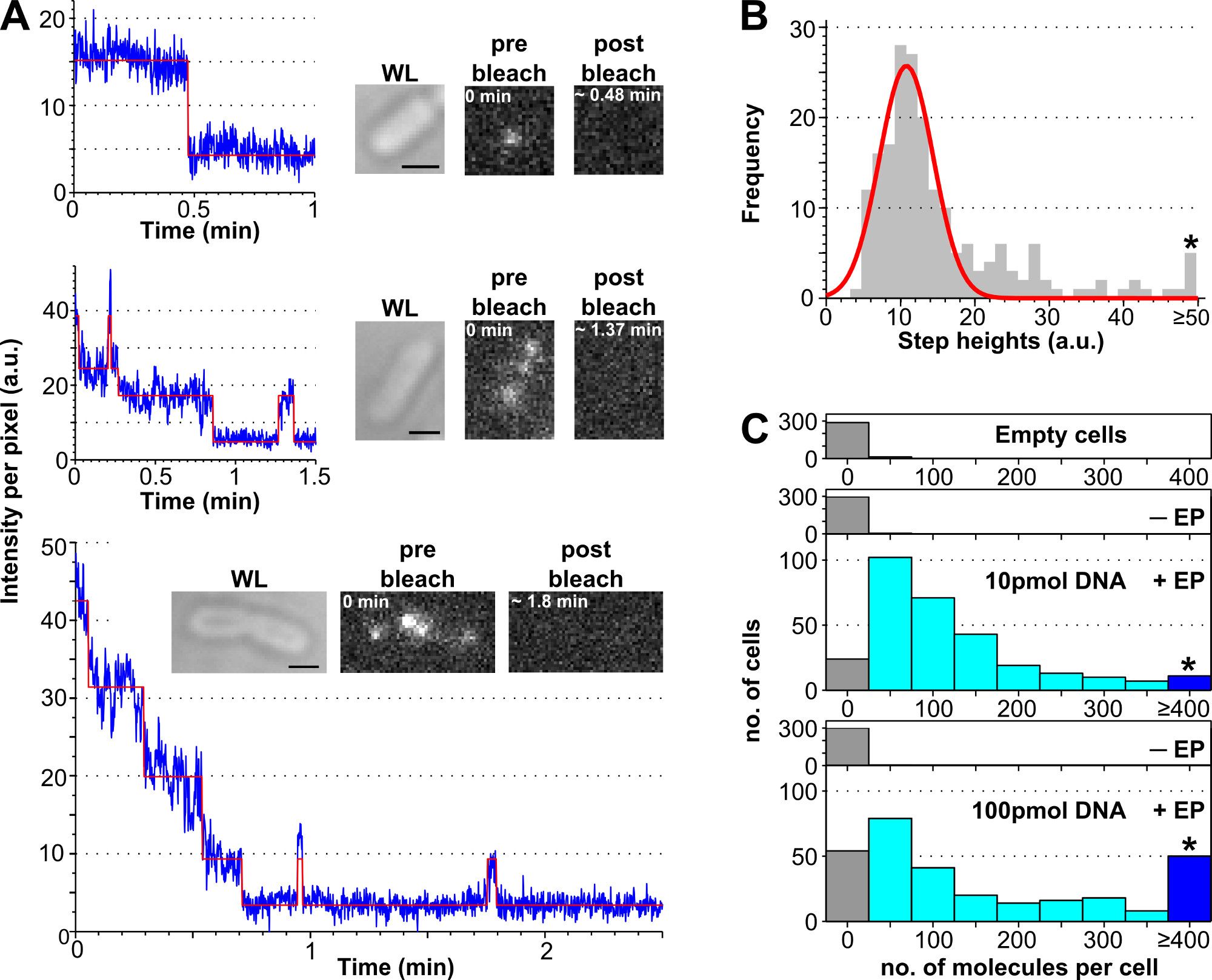
Figure 1: Counting the number of internalized molecules using photobleaching analysis.
Access restricted. Please log in or start a trial to view this content.
Materials
| Name | Company | Catalog Number | Comments |
|---|---|---|---|
| ElectroMax DH5-alpha Comptent cells | Invitrogen | 11319-019 | or any other commercial or lab-mage electrocompetant bacteria or yeast. |
| EZ Rich Defined Madia | Teknova | M2105 | low fluorescence rich media |
| MicroPulser Electroporation Apparatus | Biorad | 165-2100 | or any classical electroporator for microorganism transformation |
| Certified Molecular Biology agarose | Biorad | 161-3100 | low fluorescence agarose for agarose pad |
| Microscope coverslips No 1.5 thickness | Menzel | BB024060SC | remove background particles by heating slides in furnace at 500 °C for 1h |
| Single-molecule fluorescence microscope | Home-built | described in REFs | |
| Localization software | Custom-written, available online | MATLAB and C++ software package that can be adapted for localization analysis. | |
| Tracking software | Available online | MATLAB implementation by Blair and Dufresne. |
