This video demonstrates a fluorescence-based assay to quantify intestinal permeability in C. elegans exposed to Pseudomonas aeruginosa. Compared to controls fed Escherichia coli, pathogen-treated worms show increased fluorophore leakage into the body cavity, indicating epithelial barrier damage.
A subscription to JoVE is required to view this content. Sign in or start your free trial.
Method Article
Assessment of Intestinal Permeability in C. elegans Following Bacterial Exposure
256 views
⸱
March 31st, 2026
In This Article
Abstract
Protocol
1. Preparation of Nematode Growth Medium (NGM) Plates for Testing the Effects of Different Bacteria on the Intestinal Permeability of Caenorhabditis elegans (C. elegans)
- Place 1.25 g of peptone, 1.5 g of NaCl, 8.5 g of agar, a magnetic stirrer, and 487.5 mL of distilled water in a 500 mL glass bottle.
- Mix the mixture well, autoclave the mixture for 15 min at 121 °C, and cool the mixture to 55 °C in a water bath for 30 min.
- Remove the medium from the water bath, add the components (0.5 mL of 1 M CaCl2, 0.5 mL of cholesterol, 0.5 mL of MgSO4, 12.5 mL of KPO4) to the NGM, and mix well.
NOTE: All the individual components must be autoclaved except for cholesterol (dissolved in absolute ethanol), and every experimental step must be carried out on a clean bench. - Distribute 20 mL of NGM to each 90 x 15 mm Petri dish and allow the agar to solidify on a clean bench at room temperature (approximately 20 °C). The NGM plates can be stored for up to one month at 4 °C.
NOTE: The protocol can be paused here. The NGM plates used for FITC-dextran (Fluorescein isothiocyanate-dextran) staining were prepared. 10 mL of NGM was distributed to each 60 x 15 mm Petri dish and allowed to solidify on a clean bench at room temperature (approximately 20 °C). - Remove the bacterial culture from the 4 °C refrigerator and vortex the culture thoroughly before spreading the culture onto the NGM plates.
- Add a total of 800 µL of the bacterial culture to each fresh NGM plate, and allow the plates to dry in a 20 °C incubator overnight.
NOTE: For the first experiment (Figure 1), two NGM plates with E. coli OP50, one NGM plate with P. aeruginosa PAO1, and one NGM plate with E. faecalis KCTC3206 were prepared. For the second experiment (Figure 2), two NGM plates with live E. coli OP50, one NGM plate with live P. aeruginosa PAO1, and one NGM plate with heat-inactivated P. aeruginosa PAO1 were prepared.
3. Preparation of Age-synchronized C. elegans
- Grow worms on a solid NGM plate and feed them with E. coli OP50 until the desired population is reached.
- Synchronize the eggs by using either the timed egg laying method or the bleaching solution treatment
4. Treatment of Bacteria and FITC-dextran Feeding
- Incubate the age-synchronized eggs at 20 °C for 64 h on NGM plates supplemented with live E. coli OP50 as food.
- Wash the age-synchronized L4 larvae with an S-buffer and transfer them (more than approximately 500 worms) to the treatment NGM plates containing different bacteria and chemicals. Incubate them at 20 °C for 48 h.
NOTE: The treatment time can range from 24 to 72 h, according to the bacteria and chemicals used. The therapeutic or preventive effect of DIM can also be evaluated by pretreating the worms with pathogens and then treating the worms with DIM (the therapeutic effect) or by pretreating the worms with DIM and then treating with pathogens (the preventive effect). - To prepare the FITC-dextran-supplemented plates, mix 2 mL of heat-inactivated E. coli OP50 with 4 mg of FITC-dextran. Then, divide 100 µL of the FITC-dextran and E. coli OP50 mixture into twenty fresh NGM agar plates (60 mm x 15 mm) and allow the plates to dry for 1 h on a clean bench.
NOTE: The final concentration of FITC-dextran in each NGM plate will be 20 μg/mL. - Prepare five E. coli OP50-containing NGM plates without FITC-dextran for the vehicle control treatment. For this purpose, divide 100 µL heat-inactivated E. coli OP50 to each fresh NGM agar plate (60 mm x 15 mm) and allow the plates to dry for 1 h on a clean bench.
NOTE: For each independent experiment, fifteen FITC-dextran-supplemented NGM plates and five NGM plates without FITC-dextran are required. - After 48 h of treatment (from step 7.2), wash the worms with S-buffer, transfer the worms to the FITC-dextran supplemented plates and the NGM plates without FITC-dextran, and incubate the plates overnight (14–15 h).
NOTE: For each treatment group, 5 replicates of FITC-dextran staining (or vehicle control feeding) are required. - Wash the worms with S-buffer and allow them to crawl in the fresh NGM agar plate for 1 h.
NOTE: For each independent experiment, a total of 20 fresh NGM plates are needed. In this step, you can use the NGM plate supplemented with or without E. coli OP50. - Add 50 µL of 4% formaldehyde solution to each well of a black 96-well flat-bottom plate. Transfer approximately 50 worms from each NGM plate into each well for fluorescence measurements. After 1 to 2 min, thoroughly remove all the formaldehyde from each well and add 100 µL of mounting medium to coat the wells.
NOTE: Formaldehyde solution (4%) is used to immobilize and fix the worms. For each treatment group, 5 wells (5 replicates) are used for image analysis.
5. Imaging C. elegans with the Operetta Imaging System and Determination of Intestinal Permeability by Measuring the FITC-dextran Fluorescence Uptake
NOTE: Fluorescent stereomicroscopy can be used for image analysis instead of the Operetta system.
- Capture fluorescence images and measure the fluorescence intensity using the Operetta High-Content Imaging System and analyze the images with Harmony software.
- In the Harmony software, press the icon Open lid to open the lid and to put the plate into the machine.
- Set up the parameters.
- Click Set up, select the plate type (96-well Corning flat-bottom), and add the channels (bright-field and EGFP channels).
- Adjust the layout. Go to the Layout selection, and then select Track and adjust the parameters (first picture at 1 µm, number of planes are 10, distance is 1 µm).
- Select one of the treatment wells and one capture field and press Test to check whether the pictures are satisfactory in the Run experiment section.
- If the pictures are satisfactory, return to the Set up section and press the Reset icon at the end of the screen. Then, select all the target wells and a suitable number of capture fields.
- Go to Run experiment again and enter the plate name, then press Start to begin processing.
- To measure the intensity of the fluorescence, go to the image analysis section and input the image. Find the cell by choosing the EGFP (Enhanced green fluorescent protein) channel and method B. Adjust the common threshold to 0.5, area to >200 µm2, split factor to 3.0, individual threshold to 0.18, and contrast to more than 0.18. Then, calculate the intensity properties and choose the mean as the output. Press the Apply icon to save the setup.
- Go to the Evaluation section to measure the intensity by obtaining a heatmap and data table. Set the Readout parameter to Cells — Intensity Cell EGFP Mean — Mean per Well and start the evaluation.
NOTE: The protocol can be paused here. - To extract the data from Operetta, click the Setting button and choose Data management.
- Choose Write archive, and then open the browse and select the file.
- To select the file, click the small + signal at the left corner, and then click Measurement and choose Plate name.
- Select the file and click OK.
- Select the path to save the file by clicking the Browse signal at the active path.
- Click Start to save the data file.
NOTE: The protocol can be paused here.
Access restricted. Please log in or start a trial to view this content.
Results
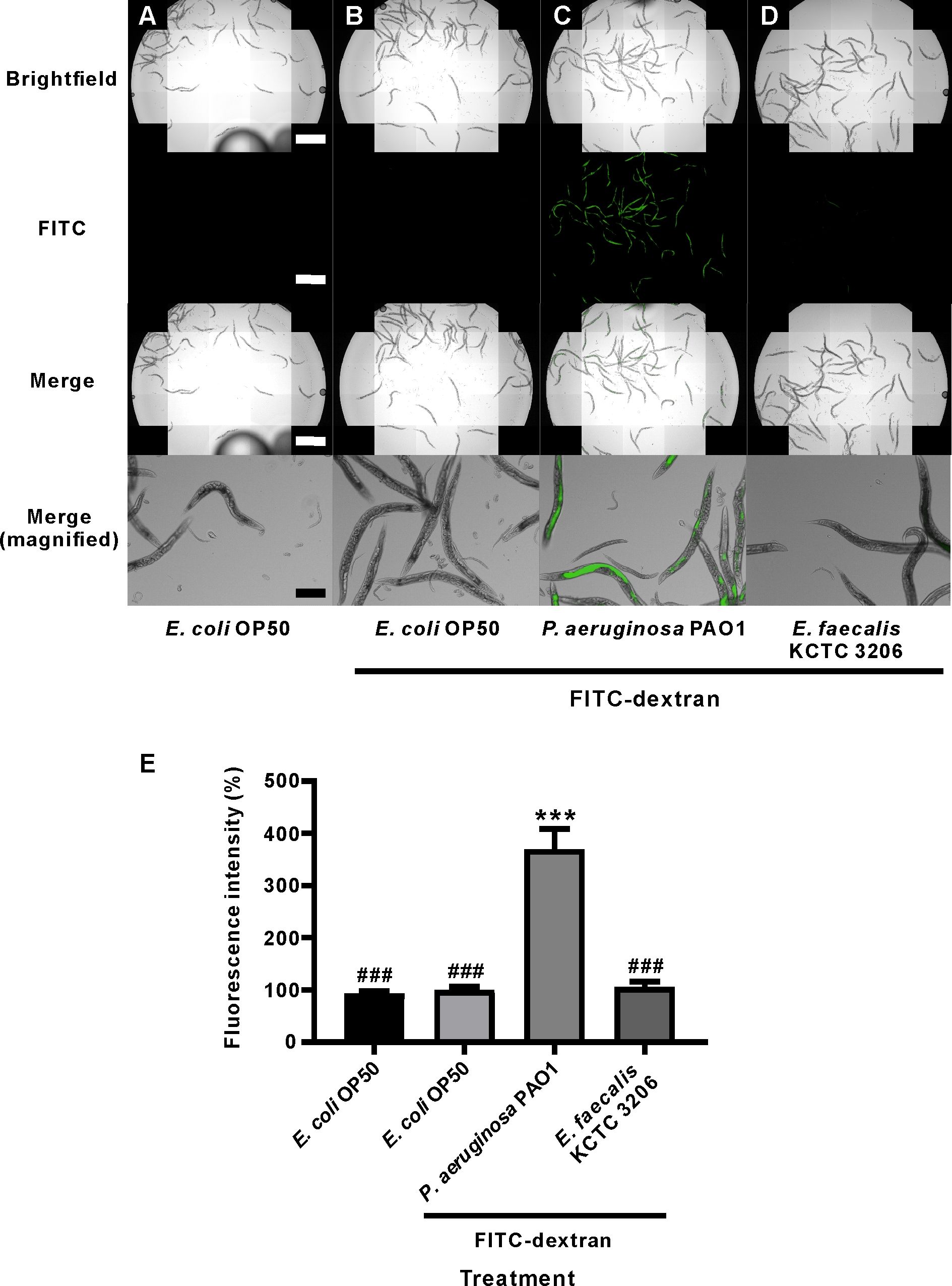
Figure 1: The effects of different bacteria on the intestinal permeability of C. elegans.
Access restricted. Please log in or start a trial to view this content.
Materials
| Name | Company | Catalog Number | Comments |
|---|---|---|---|
| 90×15 mm Petri dishes | SPL Life Sciences, South Korea | 10090 | |
| 60×15 mm Petri dishes | SPL Life Sciences, South Korea | 10060 | |
| Bactor Agar | Beckton Dickinson | REF. 214010 | |
| Formaldehyde solution | Sigma | F1635 | |
| Brain Heart Infusion (BHI) | Becton Dickinson | REF. 237500 | |
| Caenorhabditis elegans N2 | Caenorhabditis Genetics Center (CGC) | Wild type | |
| Costa Assay Plate, 96 Well Black With Clear Flat Bottom Non-treated, No Lid Polystyrene | Corning Incorporated | REF. 3631 | |
| Escherichia coli OP50 | Caenorhabditis Genetics Center (CGC) | ||
| Fluorescein isothiocyanate - dextran | Sigma | FD10S | |
| Harmony software | PerkinElmer | Verson 3.5 | |
| Luria-Bertani LB medium | Merck | VM743185 626 1.10285.5000 | |
| Magnesium sulfate heptahydrate | Fisher Bioreagents | BP2213-1 | |
| Fluoromount aqueous mounting medium | Sigma | F4680 | |
| Operetta CLS High-Content Analysis System | PerkinElmer | HH16000000 | |
| Peptone | Merck | EMD 1.07213.1000 | |
| Pseudomonas aeruginosa PA01 | Korean Collection for Type Culture | KCTC NO. 1637 | |
| Sodium Chloride | Fisher Bioreagents | BP358-1 | |
| Stereo Microscope | Nikon, Japan | SMZ800N | |
| Yeast extract | Becton Dickinson | REF. 212750 |
