This video demonstrates the preparation of spheroplasts from rod-shaped, gram-negative bacteria. Bacteria are grown in the presence of an antibiotic to form elongated filaments. These are washed with a hypertonic solution and treated with a lysis buffer to remove the outer cell wall. Hypotonic treatment forms spheroplasts, which are stabilized with a chilled isotonic solution and stored under cold conditions.
A subscription to JoVE is required to view this content. Sign in or start your free trial.
Method Article
Generation of Spheroplasts from Gram-Negative Bacteria
403 views
⸱
February 26th, 2026
In This Article
Abstract
Protocol
1. Solution Preparation
NOTE: Prepare solutions described in steps 1.1–1.9 in order to produce Escherichia coli (E. coli) spheroplasts
- Prepare 1 M tris(hydroxymethyl)aminomethane hydrochloride (Tris-Cl), pH 7.8 by dissolving 10.34 g Tris·HCl and 4.17 g of tris(hydroxymethyl)aminomethane (Tris·OH) in 50 mL of distilled water (dH₂O) in a 125 mL flask. Sterilize by filtering through a 25 mm syringe filter with a 0.2 µm membrane and store in a conical tube at room temperature.
- Prepare solution A (20 mM magnesium chloride (MgCl₂), 0.7 M sucrose, 10 mM Tris-Cl, pH 7.8) by dissolving 0.10 g MgCl₂ (95.2 g/mol) and 11.98 g sucrose (342.3 g/mol) in 25 mL dH₂O in a 125 mL flask. Add 500 µL 1 M Tris-Cl (pH 7.8) and adjust the volume to 50 mL. Sterilize by filtering through a 25 mm syringe filter with a 0.2 µm membrane and store in a conical tube at room temperature.
- Prepare solution B (10 mM MgCl₂, 0.8 M sucrose, 10 mM Tris-Cl, pH 7.8) by dissolving 0.05 g MgCl₂ (95.2 g/mol) and 13.69 g sucrose (342.3 g/mol) in 25 mL dH₂O in a 125 mL flask. Add 500 µL 1 M Tris-Cl (pH 7.8) and adjust the volume to 50 mL. Sterilize by filtering through a 25 mm syringe filter with a 0.2 µm membrane and store in a conical tube at room temperature.
- Prepare 0.8 M sucrose by dissolving 13.69 g sucrose (342.3 g/mol) in 50 mL dH₂O in a 125 mL flask. Sterilize by filtering through a 25 mm syringe filter with a 0.2 µm membrane and store in a conical tube at room temperature.
- Prepare 5 mg/mL deoxyribonuclease I (DNase I) by dissolving 0.015 g DNase I in 3 mL of dH₂O in a 50 mL flask. Sterilize by filtering through a 25 mm syringe filter with a 0.2 µm membrane. Aliquot solution into microfuge tubes and store at −20 °C.
- Prepare 0.125 M ethylenediaminetetraacetic acid (EDTA), pH 8.0, by dissolving 0.698 g EDTA disodium dihydrate (372.2 g/mol) into 15 mL dH₂O in a 125 mL flask. Sterilize by filtering through a 25 mm syringe filter with a 0.2 µm membrane and store in a conical tube at room temperature.
- Prepare 600 µg/mL cephalexin by dissolving 0.03 g of cephalexin hydrate (365.404 g/mol) in 50 mL of dH₂O in a 125 mL flask. Sterilize by filtering through a 25 mm syringe filter with a 0.2 µm membrane and store in a conical tube at 4 °C.
- Prepare 5 mg/mL lysozyme by dissolving 0.015 g lysozyme in 3 mL of dH₂O in a 50 mL flask. Sterilize by filtering through a 25 mm syringe filter with a 0.2 µm membrane. Aliquot solution into microfuge tubes and store at −20 °C.
- Prepare 3% w/v Trypticase Soy Broth (TSB) by dissolving 30 g of TSB in 1 L of dH₂O in a 2 L flask. Aliquot the solution into flasks containing 25 mL or 100 mL TSB to be used in the preparation of E. coli spheroplasts or B. megaterium protoplasts, respectively. Autoclave flasks to sterilize the liquid medium. Sterile TSB can be stored at either room temperature or 4 °C.
- Prepare solution C (1 M sucrose, 0.04 M maleate, 0.04 M MgCl₂, pH 6.5) by dissolving 34.23 g sucrose (342.3 g/mol), 0.46 g maleic acid (116.07 g/mol), and 0.38 g MgCl₂ (95.21 g/mol) in 100 mL of dH₂O in a 250 mL flask. Adjust pH to 6.5. Sterilize by filtering through a 25 mm syringe filter with a 0.2 µm membrane and store in a conical tube at room temperature.
2. Preparation of Overnight Culture
NOTE: Perform sections 2-3 using appropriate sterile techniques. If desired, a bacterial strain can contain a plasmid for antibiotic resistance to reduce the potential contamination. If using a strain with antibiotic resistance, add the necessary antibiotics in steps 2.1 and 3.1–3.2.
- Prepare an overnight culture by picking a single colony of bacteria using a sterile pipette tip and placing it into a 14 mL culture tube containing 2–3 mL of 3% w/v TSB. Incubate at 37 °C, while shaking for 16–21 h.
3. Preparation of Gram-negative E. coli Spheroplasts
- In a 250 mL flask, dilute the overnight culture 1:100 in 25 mL volume of 3% w/v TSB and incubate the bacterial solution at 37 °C, while shaking for approximately 2.5 h until the solution has reached an optical density of 0.5–0.8 at 600 nm. Measure the optical density using a spectrophotometer.
- In a 250 mL flask, dilute this culture 1:10 in 30 mL of 3% w/v TSB and incubate at 37 °C, while shaking for 2.5 h in the presence of 60 µg/mL cephalexin (347.4 g/mol) to produce single cell filaments of about 50–150 µm in length, which are observable under 1,000X magnification using a light microscope (Figure 1B).
- Harvest the filaments by centrifuging the bacterial solution at 1,500 x g, 4 °C for 4 min. Decant and discard the supernatant, reserving the pellet.
- Wash the filaments by gently adding 1 mL of 0.8 M sucrose, being careful not to disturb the pellet. Incubate for 1 min, and then discard the supernatant without disturbing the pellet.
- Add 150 µL of 1 M Tris-Cl (pH 7.8), 120 µL of 5 mg/mL lysozyme, 30 µL of 5 mg/mL DNase I, and 120 µL of 0.125 M EDTA in their respective order to the pellet and incubate the solution at room temperature for 10 min.
- Add 1 mL of solution A gradually over 1 min to the solution prepared in 3.5 using a micropipette while gently swirling the solution by hand. Incubate the solution for 4 min at room temperature.
- Put 7 mL of 4 °C solution B into two 15 mL conical tubes. Add equal amounts of the solution prepared in 3.6 to each of these two tubes. Centrifuge the solution at 1,500 x g, 4 °C for 4 min.
- Using a serological pipette, carefully remove all but 1-2 mL of supernatant without disturbing the pellet. Resuspend the pellet by gently pipetting up and down using a P1000 micropipette. Visually check to see spheroplasts formation by observing the sample at 1,000X magnification using a light microscope (Figure 1C).
- Store the spheroplasts at -20 °C for up to a week or until they have gone through 3 freeze-thaw cycles.
Access restricted. Please log in or start a trial to view this content.
Results
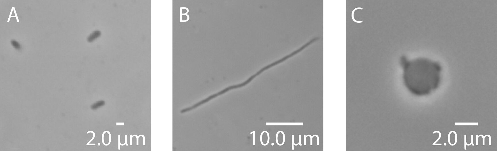
Figure 1: Representative images of Escherichia coli. (A) E. coli bacteria, (B) E. coli snake, and (C) E. coli spheroplast. Images were taken at 100X magnification.
Access restricted. Please log in or start a trial to view this content.
Materials
| Name | Company | Catalog Number | Comments |
|---|---|---|---|
| Trizma hydrocloride (Tris HCl) | Sigma | T3253 | |
| Trizma base (Tris OH) | Sigma | T1503 | |
| Magnesium chloride | Sigma | M8266 | |
| Sucrose | Sigma | S7903 | |
| Lysozyme | Sigma | L6876 | |
| Deoxyribonuclease I | Sigma | D4527 | |
| Ethylenediaminetetraacetic acid | Sigma | 106361 | Used Sigma 106361 in original protocol development; 106361 discontinued with ED2SS as replacement |
| Cephalexin hydrate | Sigma | C4895 | |
| BBL Trypticase soy broth | Fisher Scientific | B11768 | |
| Maleic acid | Sigma | M0375 | |
| Acrodisc 25 mm Syringe Filter w/ 0.2 μm HT Tuffryn Membrane | Pall Corporation | 4192 |
