This video demonstrates the process of quantifying bacterial load in the spleen of an infected mouse to assess the severity of infection. The homogenized spleen is serially diluted and plated to determine total bacterial burden. The resulting data provide insights into host-pathogen interactions within systemic tissue.
A subscription to JoVE is required to view this content. Sign in or start your free trial.
Method Article
Assessing Bacterial Burden in the Mouse Spleen for Infection Studies
280 views
⸱
February 26th, 2026
In This Article
Abstract
Protocol
All procedures involving animal models have been reviewed by the local institutional animal care committee and the JoVE veterinary review board.
1. Murine Intratracheal Aspiration of Klebsiella pneumoniae from the Oropharynx
- Ensure that mice are uniquely identified by tail mark, tattoo, or other approved identifier.
- Draw up a dosing inoculum of bacteria using a P200 pipette prior to removing mice from the isoflurane in order to expedite the procedure. For best results with i.t. aspiration, use a volume of <100 µl to prevent overflow. Herein, use 2000 colony-forming units (CFU)/50 µl of K. pneumoniae.
- Anesthetize mice in a clear chamber with isoflurane gas (e.g., 2% isoflurane at flow rate of 1 L/min) or as per institutional guidelines. The number of mice to anesthetize together is determined by the comfort level of the experimenter, typically 1-2 at a time. Observe breathing and confirm level of anesthesia once deep breaths are visible and 2-3 sec can be counted between breaths.NOTE: If there is concern about possible confounding effects of volatile (inhaled) anesthetics, anesthesia can be achieved instead with intraperitoneal injection of ketamine/xylazine. With the method described above, deep anesthesia only lasts a few minutes. If more prolonged anesthesia is induced, eye ointment should be used to prevent ocular desiccation.
- Once the mouse is anesthetized, position it in a semirecumbent supine position, suspended by the maxillary incisors from a rubber band stretched between pegs on a slanted Plexiglas board (Figure 1).
- Gently pull the mouse tongue to the side with a pair of blunt, non-ridged forceps and deposit the dose into the oral cavity with a P200 pipette. Exercise great care to avoid inducing trauma to either the tongue or the oropharynx.
- While keeping the tongue retracted, gently occlude the nose with a gloved finger until the mouse inhales. Continue to cover the nose until the mouse has taken two or more inhalations, and no liquid is visible in the oral cavity. Covering the nose helps ensure that the mouse will inhale the bacteria into the lungs, as mice are obligate nose breathers.
- Remove the mouse from the inoculation board and return to its cage, placing the mouse on its back to prevent bedding or debris from blocking the nares while the mouse is recovering from anesthesia.
- Once all mice have been dosed and have awoken from anesthesia, house mice in a cubicle/room containing only mice that have been dosed with K. pneumoniae. Monitor mice daily, including body weights, if going beyond 48 hr post-infection.
- Use daily mash and/or supplemental heat if allowing the mice to go beyond 48 hr.
2. Determining Bacterial Load in Lung and Peripheral Tissues
- Before beginning: prepare dilution tubes with 900 µl of 1x phosphate-buffered saline (PBS) for lung and spleen and 90 µl of 1x PBS for blood. Label plates for culturing bacteria and set at room temperature; therefore, once tissue has been collected, time will be minimized between homogenization and plating for bacterial growth.
NOTE: Due to the heterogeneous deposition of bacteria in the lungs that can occur with aspiration, it is recommended that individual mice be used for analysis of either total airway cellularity or total (bilateral) lung bacterial burden. - Euthanize mice per institutional guidelines. Here, use a lethal injection of 150 mg/kg sodium pentobarbital or an equivalent dose of commercial euthanasia solution, followed by exsanguination, as this avoids possible complications to the lungs associated with CO2 inhalation and cervical dislocation.
- Remove the spleen and place it into 2 ml of 1x PBS. Place tubes on ice. Take care to clean instruments with ethanol between each tissue/mouse.
- Homogenize the tissue on ice, preferably with disposable homogenizers that can be changed between each sample. Disposable homogenizer tips can be autoclaved for reuse between experiments.
- Once tissue has been homogenized, perform serial dilutions and plate. Plate samples of a given tissue/dilution in duplicate on a single tryptic soy agar (TSA) plate by drawing a dividing line on the plastic. Plating and dilution suggestions are shown below (in all cases, 10 µl of sample is spread onto the plate) and are based on a 24 hr post-infection necropsy. If analyzing 48-72 hr samples, a larger number of dilutions may need to be plated due to increased bacterial burden at later time points
- For Spleen - dilute 1:10-1:100 by adding 100 µl of homogenized spleen into 900 µl of sterile 1x PBS serially. Plate neat homogenate and both dilutions in duplicate.
- Allow the spread samples to dry briefly.
- Invert TSA plates and place in a static 37 °C incubator overnight.
- Determine the number of colonizing bacteria by averaging the bacterial colonies from both sides of the plate and from each dilution. Factor in the initial dilutions of 5 ml for the lung and 2 ml for the spleen to determine total tissue CFUs.
Access restricted. Please log in or start a trial to view this content.
Results
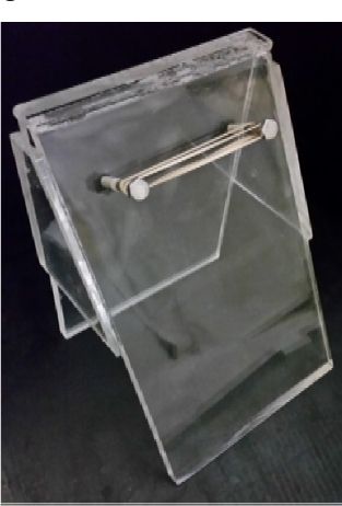
Figure 1: Procedure Board for Aspiration Pneumonia in Mice. Study mice are positioned, hea...
Access restricted. Please log in or start a trial to view this content.
Materials
| Name | Company | Catalog Number | Comments |
|---|---|---|---|
| ATCC | 43816 | ||
| Becton Dickenson | 211825 | ||
| Prepared in-house | n/a | Distilled water (5 L), NaCl (40 g), KCl (1 g), Na₂HPO₄ (5.75 g), KH₂PO₄ (1 g) |
