This video demonstrates the preparation and imaging of fluorescently labeled pathogenic bacteria. Agarose pads are first prepared and positioned within an adhesive frame. A suspension of genetically modified bacteria expressing a fluorescent protein is applied to the pad to immobilize the cells. The frame is then sealed with a coverslip and observed under a microscope to capture fluorescence images of bacteria.
A subscription to JoVE is required to view this content. Sign in or start your free trial.
Method Article
Sample Preparation and Imaging of Fluorescently Labeled Bacteria
502 views
⸱
March 31st, 2026
In This Article
Abstract
Protocol
NOTE: All procedures involving the growth, manipulation, and imaging of L. pneumophila should be performed in a biological safety level 2 laboratory in compliance with local guidelines.
1. Insertion of sfGFP into Legionella pneumophila Chromosome Using Allelic Exchange and Double Selection Strategy (Figure 1, Figure 2)
- Clone into the gene replacement vector pSR47S11 the following sequence: the 1,000 bp upstream of the site of interest, then the sfGFP sequence, then the 1,000 bp downstream of the site of interest (Figure 1). The sfGFP sequence should be placed in frame to the N-terminus or C-terminus ends with a linker that contains four to eight amino acids. Transform the resulting vector into Escherichia coli DH5αλpir. Later, streak L. pneumophila (the recipient) for single colonies on charcoal-yeast extract (CYE) agar containing 100 µg/mL streptomycin and grow for 5 days at 37 °C (Figure 2).
- Streak L. pneumophila on CYE-agar-streptomycin and grow for 2 days at 37 °C (heavy patch). Streak E. coli DH5α transformed with pRK600 helper plasmid (helper) on LB agar containing 25 µg/mL chloramphenicol. Streak the E. coli DH5αλpir (donor) on LB agar containing 50 µg/mL kanamycin.
- Perform triparental mating: incubate a colony of the helper, a colony of the donor, and the recipient by overlaying patches of the three strains on a CYE agar plate without selection and incubating for 4–8 h at 37 °C. As negative controls, incubate a helper+recipient strain mix and a donor+recipient strain mix for the same periods of time.
- Resuspend the mating reactions in 500 µL of ddH2O. Plate 20 µL and 50 µL of the reactions on CYE agar containing 100 µg/mL streptomycin and 10 µg/mL kanamycin and grow for 5 days at 37 °C. Streak four of the resulting clones on CYE agar containing 100 µg/mL streptomycin and grow for 5 days at 37 °C.
- Streak 16 clones on CYE agar containing 5% sucrose and 100 µg/mL streptomycin and grow for 5 days at 37 °C. Then, streak 32 of these clones on CYE agar containing 100 µg/mL streptomycin and on CYE agar containing 100 µg/mL streptomycin and 10 µg/mL kanamycin and grow for 5 days at 37 °C.
2. Isolation of Clones that Integrated sfGFP into the L. pneumophila Chromosome
- Streak clones that were sensitive to kanamycin on CYE-agar-streptomycin plates confirm the insertion of sfGFP into the chromosome with colony polymerase chain reaction (PCR). Use primers that are complementary to the sfGFP gene and to the chromosomal region of interest to amplify the insertion junction.
- Mix 0.5 µL of each of the 10 µM primer solutions and one colony to a final volume of 12.5 µL and denature for 10 min at 95 °C. Cool on ice for 10 min, add 12.5 µL of 2x PCR master mix solution, and perform a PCR analysis.
- Grow heavy patches of the isolated colonies on CYE-agar-streptomycin plates for 2 days at 37 °C. Examine the expression levels and stability of the sfGFP fusions by immunoblotting with an anti-GFP antibody.
3. Live Cell Imaging of L. pneumophila with Fluorescently Tagged Dot/Icm Components
- Preparation of agarose pads
- Make about 30 mL of a 1% low-melt agarose solution in water. Microwave in a glass flask for about 90 s, swirling occasionally, until the agarose is completely dissolved.
- Place two 22 x 22 x 0.15 mm3 glass slides on the edge of a 25 x 75 x 1.1 mm3 glass slide, one on top of the other. Stack two more 22 x 22 x 0.15 mm3 glass slides on the other edge.
- Pipette about 1 mL of the molten agarose into the center slide between the two upper glass slides, then place another 25 x 75 x 1.1 mm3 slide on top of the molten agarose. Try to avoid the formation of air bubbles. Cool the slides at 4 °C for 15 min.
- Using a scalpel or razor blade, gently cut the pad into small squares, ~5 x 5 mm2. Fix a double-sided adhesive 17 x 28 x 0.25 mm3 frame on a 25 x 75 x 1.1 mm3 glass slide and place several pads on the slide.
- Image acquisition
NOTE: The following steps are described for a microscope that is under the control of SlideBook 6.0 and equipped with solid state illuminators, CCD monochrome camera, and a 100x objective lens (1.4 numerical aperture). If needed, use alternative microscopy devices with appropriate hardware and software configurations that can be customized according to the protocol settings.- Dissolve a heavy patch of L. pneumophila in 1 mL of ddH2O, vortex and pipet 2–3 µL of the dilution onto the pads. Place a 50 x 24 x 0.15 mm3 coverslip gently over the adhesive frame.
- In the capture window adjust the ND to 180. Adjust the binning to 2×2 and use the 488 nm channel to expose the sample between 500–1,000 ms. Validate the specificity of the fluorescence signal by imaging untagged L. pneumophila with the same parameters (Figure 3).
Access restricted. Please log in or start a trial to view this content.
Results
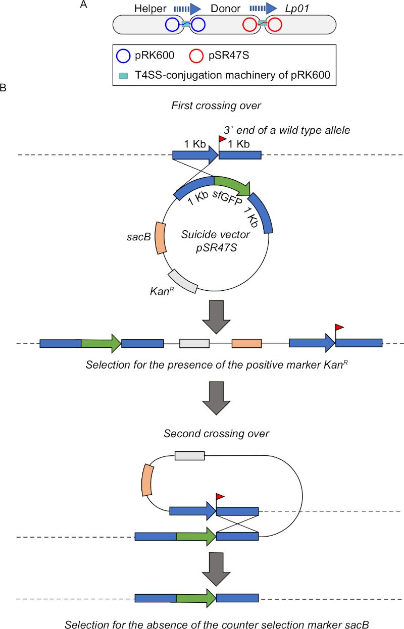
Figure 1: Schematic overview of homologous recombination and allelic exchange used to insert sfGFP into
Access restricted. Please log in or start a trial to view this content.
Materials
| Name | Company | Catalog Number | Comments |
|---|---|---|---|
| 100x Plan Apo objective (1.4 NA) | Nikon | ||
| ACES | Sigma-Aldrich | A9758 | |
| Activated charcoal | Sigma-Aldrich | C5510 | |
| Agarose GPG/LMP, low melt | American bioanalytical | AB00981 | |
| Bacto dehydrated agar | BD | 214010 | |
| Gene Frame, 1.7x2.8 cm, 125 µL | Fisher Scientific | AB-0578 | |
| L-Cysteine | Sigma-Aldrich | C7352 | |
| Microscope cover slides 22x22 mm | Fisher Scientific | 12-542B | |
| Microscope cover slides 24x50 mm | Fisher Scientific | 12-545K | |
| Microscope slides 25x75x1 mm | Globe Scientific | 1380 | |
| SlideBook 6.0 | Intelligent Imaging Innovations | ||
| Spectra X light engine | Lumencor | ||
| Taq 2X Master Mix | New England BioLabs | M0270 | |
| Titan Krios | Thermo Fisher Scientific | ||
| Yeast Extract | BD | 212750 |
