This video demonstrates the procedure for establishing a bacterial infection in Drosophila larvae by placing them into a bacterial droplet and pricking them with a tungsten needle. The larvae are then incubated on a grooved, nutrient-rich agar plate to study host-pathogen interactions.
A subscription to JoVE is required to view this content. Sign in or start your free trial.
Method Article
Establishing Bacterial Infection in Drosophila Larvae
352 views
⸱
March 31st, 2026
In This Article
Abstract
Protocol
1. In vivo infection
- Warm the Drosophila fruit juice agar plate at room temperature for 15 min.
- Add 30 g of agar to 700 mL of water and autoclave it for 40 min.
- Dissolve 0.5 g of methyl paraben in 10 mL of absolute ethanol.
- Add the methyl paraben solution to 300 mL of fruit juice concentrate.
- Quickly mix the juice concentrate into the autoclaved agar solution and dispense 5 mL into 10 × 35 mm Petri dishes.
- After the plates have cooled for 15 min, store them at 4 °C.
- Prepare 3rd instar larvae.
- Place the yeast paste on an agar plate. Make a fine cut in the agar plate where larvae can migrate to avoid drying (Figure 1A). Assemble a 0.001 mm pointed tungsten needle with holding forceps using paraffin film (Figure 1B, C).
- Pipette 50 µL of high-titer mCherry expressing-Coxiella burnetii (5.95×10⁹ GE/mL) onto the paraffin film under the stereo microscope and place the larvae into the pool of bacteria.
- Place the larvae in the pool of bacteria, prick them with a tungsten needle (Figure 3D), and transfer them onto an agar plate (Figure 1E).
- Transfer the remaining pathogen medium onto the agar plate and seal the plate with paraffin film.
- Keep the larvae on the plate in moist air until the desired time post-infection (Figure 1F). In this experiment, C. burnetii-infected larvae are on the plate for 24 h.
Access restricted. Please log in or start a trial to view this content.
Results
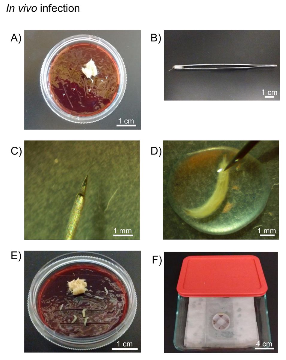
Figure 1: In vivo infection. A) Drosophila fruit juice agar plates and yeast paste...
Access restricted. Please log in or start a trial to view this content.
Materials
| Name | Company | Catalog Number | Comments |
|---|---|---|---|
| Stereo microscope | Amscope | SM-1BSZ-L6W | |
| Forceps | VWR | 82027-402 | |
| Coxiella burnetii - mCherry | Dr. Heinzen, R. | ||
| Drosophila fruit juice plates | Cold Spring Harbor Protocols | http://cshprotocols.cshlp.org/content/2007/9/pdb.rec11113.full | |
| Agar | Fisher Bioreagents | BP1423-500 | |
| Methyl paraben | Amresco | 0572-500G | |
| Absolute ethanol | Fisher Bioreagents | BP2818-500 | |
| Welch's 100% Grape juice frozen concentrate, 340 mL | Amazon | B0025UJVGM | |
| Petri dishes, 10 x 35 mm | Fisher Scientific | 08-757-100A | |
| Yeast, Bakers Dried Active | MP Biomedicals | 210140001 | Add 2 parts of water to 1 part of yeast (v/v) |
| Tungsten needle | Fine Science Tools | 10130-20 | |
| Holding forceps | VWR | HS8313 | |
| Listeria monocytogenes | ATCC | strain: 10403S | Listeria monocytogenes strain 10403S (Bishop and Hinrichs, 1987) was grown in Difco Brain-heart infusion (BHI) broth (BD Biosciences) containing 50 µg/ml streptomycin at 30 °C. |
