The video demonstrates bacterial load measurement in spleen and liver tissues isolated from mice infected with an intracellular bacterial pathogen. Bacteria, accumulated within tissue-resident phagocytes during systemic infection, are released using detergent-based lysis and mechanical disruption. Serial dilutions of the homogenates are plated on nutrient agar, and following incubation, colony counts indicate the bacterial burden within each organ.
A subscription to JoVE is required to view this content. Sign in or start your free trial.
Method Article
Quantification of Bacterial Load in Mouse Spleen and Liver After Systemic Infection
205 views
⸱
July 1st, 2026
In This Article
Abstract
Protocol
Disclaimer Text: All procedures involving animal models have been reviewed by the local institutional animal care committee and the JoVE veterinary review board.
1. Preparation of Glycerol Stocks for Long-term Storage
NOTE: This procedure describes how glycerol stocks of the EGD strain of L. monocytogenes are prepared from an original glycerol stock. Steps that have the potential to generate aerosols should be performed within a certified biosafety cabinet (BSC).
- Prepare brain heart infusion (BHI) agar plates for bacterial growth. For this, add 3.8% (w/v) BHI broth and 1.5% (w/v) agar to double-distilled water (ddH2O). Autoclave liquid. Once the agar cools to 50 °C, dispense liquid into bacterial petri dishes (25 mL/dish) and let plates dry (uncovered) in the BSC for 1 hr.
NOTE: Transfer BHI agar into a 50 °C water bath after autoclaving to avoid solidification prior to pouring plates. Store BHI plates at 4 °C upside down (with media side on top) until ready for use. - Prepare liquid BHI media. For this, mix 3.8% (w/v) BHI broth in ddH2O. Autoclave.
- Remove frozen glycerol stock of the L. monocytogenes EGD strain from the -80 °C freezer and thaw to room temperature.
- Dip a sterile pipette tip in the thawed glycerol stock and immediately streak the tip back and forth across a section of a BHI plate. This is the primary streak.
- Turn the plate by 90 °C and using a fresh pipette tip, drag through the first streak and spread it to the next ¼ of the plate (this is the secondary streak). Repeat once more to make the tertiary streak.
- Turn the plate upside down and incubate at 37 °C overnight. Single uniform colonies should be obtained in the last set of streaks and visible between 16 and 24 hr.
- Dispense 10 mL of sterile BHI broth into a sterile vented 50 mL tube. Pick one colony of L. monocytogenes from the plate using a sterile pipette tip and inoculate the broth. Incubate the culture in a 37 °C orbital shaking incubator overnight or until OD600 = 1.0 with settings at 225 rotations per min (rpm).
NOTE: Glass or disposable plastic Erlenmeyer flasks can also be used to culture bacteria. Regardless of the type of container used, make sure that it is sterile, vented, and that the volume of culture does not exceed 20% of the total volume of the container to ensure appropriate aeration of the bacteria. - Prepare glycerol stocks by mixing sterile 100% glycerol with an overnight bacterial liquid culture at a 1:1 ratio. Distribute the bacterial/glycerol mixture into 2 mL cryogenic vials (500 µL/vial) and transfer vials to -80 °C freezer for storage.
NOTE: Bead stock methods can also be used in place of glycerol stocks to store bacteria. By this method, porous microbeads are inoculated with a pure culture of L. monocytogenes and are stored at -80 °C. Each bead can be used to inoculate a fresh culture as needed. See materials list for further information.
2. Determination of Growth Curve of L. monocytogenes in Day Culture
NOTE: This procedure describes how to generate the growth curve for L. monocytogenes that is used to estimate the colony-forming units (CFU) for infection studies. All steps that have the potential to generate aerosols should be performed within a certified BSC.
- Take 100 µL of the overnight culture generated in Step 1.7 to 10 mL BHI media in a vented 50 mL tube and grow at 37 °C in a shaking incubator (225 rpm, tilted at a 45° angle). Use a non-inoculated tube as a control.
- Take 0.5 mL samples of the culture at hourly intervals (1, 2, 3, 4, 5, 6 hr, etc.). Dilute each aliquot 1:1 (v/v) with BHI media in a plastic cuvette. Pipette up and down to mix. Measure the optical density (OD) at 600 nm (OD600) using a spectrometer. Continue culturing bacteria until OD600=1.
- At the same time, take a 100 µL sample of the culture and dilute with 900 µL BHI media in a sterile 1.5 mL microcentrifuge tube (this is the 10-1 dilution). Centrifuge the bacteria at 6,000 x g for 5 min, and aspirate the supernatant.
- Wash bacteria twice by resuspending the pellet in 1 mL of BHI media, centrifuging for 5 min at 6,000 x g, and then aspirating the supernatant. Resuspend the pellet in 1 mL BHI media. Prepare a 10-fold dilution series of this sample in BHI media (10-2 to 10-9). Spread 100 µL of each diluent onto separate BHI agar plates. Incubate plates overnight at 37 °C in an incubator.
- The next day, pick plates that have between 30 - 300 colonies. Discard the rest. Count the colonies on these plates. Table 1 shows an example of counts obtained in an aliquot that was taken when OD600 = 0.84. In this example, one of the plates (i.e., 10-6 dilution) had colony counts between 30 - 300 and was used for the CFU/mL calculation.
NOTE: Plates with greater than 300 colonies are not used, since overcrowding can hinder bacterial growth and also make it difficult to discern and enumerate individual colonies. Plates with counts < 30 are also not used because small errors in the dilution technique or the presence of contaminants can have a large impact on the precision of counts at the lower end of the range. - Divide the number of colonies by the volume plated and then multiply by the dilution factor to obtain the CFU/mL value for a particular dilution. In the example in Table 1, the count at the 10-6 dilution was 70. Divide this value by 0.1 mL to get the CFU/mL value for the diluted culture. Then multiply this value by the dilution factor (106) to obtain the CFU/mL value of the undiluted culture (7.0 x 10⁸).
- Plot the OD600 (y-axis) versus time in h (x-axis) to identify the logarithmic phase of growth.
NOTE: This growth curve provides an estimate of the CFU/mL of the day culture when grown to a certain OD reading. Choose an OD600 reading that is in the logarithmic phase of growth that can be used as a target OD600 for growing day cultures. These data can now be used to estimate the CFU in a culture for the preparation of inoculum (Procedure 3).
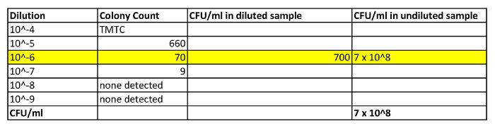
Table 1: Shows Some Representative Calculations for Determining CFU in an Aliquot of Day Culture. In this example, an aliquot of the day culture was taken and diluted 1:1 with BHI media. The OD600 of this diluted sample was determined to be 0.84. In addition, a 100 µL aliquot was taken for CFU determination. This sample was diluted with 900 µL of BHI media (10-1) and was washed and resuspended in 1 mL BHI. A 10-fold dilution series of this sample was prepared (10-2 to 10-9), and diluted samples were plated on BHI agar plates (only values for 10-4 to 10-9 are shown). The next day, the colonies were counted. Only those plates that had colony numbers between 30-300 were considered for the calculation (i.e., 10-6 plates, highlighted in yellow). The number of colonies on this plate (70) was then divided by 0.1 (volume in mL plated) to get the CFU/mL of the diluted sample. This value was then multiplied by the dilution factor (106) to obtain the CFU/mL reading of the undiluted culture. TMTC = too many to count.
3. Preparation of the Inoculum for Experimental Infection with L. monocytogenes
NOTE: This procedure describes the preparation of the infectious inoculum from a day culture that was started from an overnight culture (prepared in Procedure 2). All of these steps are performed in the BSC unless otherwise indicated.
- Calculate the number of CFU required for infection based on the number of mice and the experimental design of the study. Add an appropriate volume of BHI media to a sterile vented Erlenmeyer flask or culture tube.
NOTE: The CFU of bacteria prepared will be dependent on the type of experiment performed. If measuring bacterial load, each mouse is inoculated with 2 x 10⁴ CFU of bacteria. - Inoculate the tube containing BHI media with 100 µL of the overnight culture. Incubate the culture in a 37 °C orbital shaking incubator (225 rpm) until the target OD600 is reached. Transfer culture contents into a sterile centrifuge tube.
- Centrifuge bacteria into a pellet for 5 min at 6,000 x g using a centrifuge. Aspirate the supernatant using a vacuum attached to a trap flask containing bleach.
- Wash pellet twice with 1x phosphate-buffered saline (PBS), centrifuging (5 min at 6,000 x g) in between.
- Aspirate the second wash and dilute bacteria at the appropriate concentration in 1x PBS to deliver the CFU of interest to each mouse in a 200 µL volume.
NOTE: It is best to use a commercial source of sterile 1x PBS for washing bacteria and for preparation of the inoculum, since lab glassware can introduce immunological contaminants such as lipopolysaccharide.
4. Experimental Infection of Mice with L. monocytogenes
NOTE: This procedure describes how to infect mice with the inoculum prepared in Procedure 3 and how to verify the CFU delivered in the inoculum. Handling of mice and injections are performed in a BSC.
- Order a sufficient number of male or female C57BL/6J mice for your experiment. Also, order mice to serve as uninfected controls.
- Allow mice to acclimatize for 1 week prior to bacterial inoculation.
NOTE: This is because the stress associated with transport of the animals can trigger a transient increase in stress hormone production and lymphopenia. - On the day of inoculation, obtain a baseline body weight for each mouse and record it in the lab notebook.
- In the BSC, mix the bacterial suspension up and down using a sterile pipette to ensure that the bacteria are evenly distributed and then take up 200 µL of the inoculum into a 1 mL safety engineered syringe fitted with a 25 G needle.
- Inject a mouse intraperitoneally (i.p.) with 200 µL of prepared inoculum. For this procedure, scruff the mouse with the less dominant hand by grabbing the loose skin around the mouse's shoulders. After ensuring that the mouse is well-restrained, inject the mouse in the lower quadrant of the abdomen, just lateral to the midline, to avoid the bladder.
- Dispose of the needle and syringe in a biohazard sharps container.
- Repeat steps 4.3 - 4.6 until all mice are injected. Conduct similar steps with 1x PBS-injected mice (non-infected controls).
NOTE: Since the CFU is an estimate based on the growth curve, it is also good practice to check the actual CFU in the inoculum. For this, prepare 3 - 4 different dilutions of the prepared inoculum (using a 10-fold dilution series) that you expect will result in countable colonies. Spread 100 µL of each diluent onto a BHI agar plate and incubate overnight at 37 °C. Count the colonies and calculate the actual CFU/mL as described in Procedure 2.
5. Measurement of Bacterial Load in the Spleen and Liver at the Time of Peak Infection
NOTE: All steps are performed within a BSC unless otherwise noted.
- Inoculate mice i.p. with 2 x 10⁴ CFU of the pathogen using procedures described in Procedure 4.
- On day 3 post-infection, prepare sterile 1.5 mL microcentrifuge tubes, each containing 500 µL of ice-cold sterile 0.1% Triton X-100 in 1x PBS and 0.2 - 0.3 g of 1.5 - 2 mm acid-washed sterile glass beads. Weigh each tube.
NOTE: Glass beads are acid-washed by incubating in 10% acetic acid in a beaker on a magnetic stirrer for 1 hr. These beads are then extensively washed with ddH2O to remove acid, are air-dried, and then autoclaved prior to use. - Euthanize mice by CO2 exposure.
- For the dissection of organs, lay the animal on its back on a dissecting board and pin the limbs of the mouse to the board using 25 G needles. Disinfect skin by wetting it with 70% ethanol.
- Using sterile tough cut scissors, make a midline incision in the skin from the groin to the mid chest and then from the mid groin towards each knee and from the mid chest towards each elbow. Bluntly dissect and reflect back the skin, pinning it open using 25 G needles.
- Disinfect the muscle layer by wetting it with 70% ethanol and then using sterile fine scissors, make a midline incision in the peritoneal wall. Grab the xiphoid process with forceps. Then, using the same fine scissors, make cuts in the peritoneal wall from the xiphoid process laterally on each side, following the rib cage, just below the diaphragm to reveal the liver.
- Cut out a ~100 mg piece of the liver (use the same lobe for all mice) using sterile scissors and place it in a pre-weighed 1.5 mL microcentrifuge tube.
- Use forceps to gently push aside the organs on the left side of the peritoneal cavity to visualize the spleen. Gently grab the spleen with a pair of forceps and release it from the peritoneal cavity by cutting away the surrounding connective tissue.
- Place the spleen in the pre-weighed 1.5 mL microcentrifuge tube containing beads. Transport tissues to the laboratory in a leak-proof container containing ice. Re-weigh the tubes containing the organs to determine the tissue weights in mg.
- Homogenize the tissues by shaking the tubes using a bead mill homogenizer for 3 min at a frequency of 30 Hertz.
NOTE: The bead mill method is preferred for homogenization as it is amenable to processing a large number of samples and creates less mess and potential exposure to the pathogen. However, automatic homogenizers or autoclaved 2 mL manual glass tissue homogenizers could be used as an alternative. - Prepare a 10-fold dilution series of the homogenates in 0.1% Triton-X-100 in 1x PBS, ranging from (ranging from undiluted to 10-7).
- Spread 100 µL of each diluted homogenate onto a BHI agar plate (in duplicate) using a sterile spreader. Transfer plates to a 37 °C incubator and incubate overnight.
- Keep the plates that contain between 30 and 300 colonies/plate, and discard the rest. Count colonies on each plate and determine the mean number of colonies for duplicate spreads.
- Calculate the CFU/mg according to the following equation:CFU/mg = CFU/mL in the homogenate, multiplied by the mL of homogenate prepared, divided by the mg weight of the tissue homogenized.
NOTE: For example, if a mean of 30 colonies was counted after plating 100 µL of 10-2 diluted homogenate prepared from a 120 mg piece of liver that was homogenized in 0.5 mL, the calculations would be as follows:CFU/mL = 30 colonies x 100 (dilution factor) / 0.1 mL (volume spread) = 30,000 CFU/mL.CFU/mg = 30,000 CFU/mL x 0.5 mL homogenate / 120 mg tissue = 125 CFU/mg.
Access restricted. Please log in or start a trial to view this content.
Materials
| Name | Company | Catalog Number | Comments |
|---|---|---|---|
| Brain Heart Infusion Broth, Modified | BD | 299070 | any brand should be appropriate |
| Agar | BD | 214010 | any brand should be appropriate |
| Triton X-100 | Sigma-Aldrich | X100 | any brand should be appropriate |
| 1x PBS | Sigma | D8537 | any brand should be appropriate |
| TissueLyser II | Qiagen | 85300 | any brand should be appropriate |
| 16% Paraformaldehye | Electron Microscopy Sciences | 15710 | Dilute to 4% PFA in ddH2O or 1x PBS |
| 10x Perm/Wash buffer | BD | 554723 | Dilute 1x in ddH2O |
| OneComp eBeads | eBioscience | 01-1111 | Use one drop per stain |
| 50 ml vented tubes for culture | Used for culturing the bacteria, any brand should be appropriate | ||
| 1.5 ml microcentrifuge tubes | any brand should be appropriate | ||
| Bacterial petri dishes | any brand should be appropriate | ||
| 2 ml cyrovials | any brand should be appropriate | ||
| UV spectrometer | any brand should be appropriate | ||
| safety engineered needles | any brand should be appropriate | ||
| C57BL/6J | Jackson laboratories | Stock#000664 | Order for arrival at 7 weeks |
| 70% Ethanol | For decontamination | ||
| Glass beads | any brand should be appropriate | ||
| Centrifuge | rotor, buckets, bucket covers. | ||
| Microcentrifuge | any brand should be appropriate | ||
| Sterile Glycerol | any brand should be appropriate | ||
| Pipette Tips | any brand should be appropriate | ||
| Pipette | any brand should be appropriate | ||
| Surgical instruments | any brand should be appropriate | ||
| 3 ml syringe | any brand should be appropriate | ||
| Filtration Units | any brand should be appropriate | ||
| Microbank Bacterial Preservation System | Pro-lab Diagnostics | Used as an alternative to glycerol stocks for long-term storage of bacteria |
