This video demonstrates the method of AAV-mediated gene delivery in a transgenic mouse retina, followed by tissue dissection and staining to assess inner limiting membrane integrity.
A subscription to JoVE is required to view this content. Sign in or start your free trial.
Method Article
AAV-Mediated Gene Delivery for Visualization of Compromised Retinal Membrane Integrity
106 views
⸱
August 31st, 2026
In This Article
Abstract
Protocol
All procedures involving animal samples have been reviewed and approved by the appropriate animal ethical review committee.
1. Immunohistochemistry
- For retinal flatmounts (Figure 1), fix enucleated eyes in 4% paraformaldehyde for 15 min to facilitate the dissection.
- In 15-30 min, dissect the eyes to remove the cornea and lens. Separate the retina from the retinal pigment epithelium (RPE) and sclera by cutting around the ora serrata and the optic nerve. Immerse in 4% paraformaldehyde for another 30 min to fix the retina and the fluorescent protein in transduced retinal cells (fixation must not exceed 24 hr). Wash 5 min in sterile PBS, pH 7.4.
- Change the phosphate-buffered saline (PBS) to blocking buffer (PBS with 1% bovine serum albumin (BSA), 2% normal goat or donkey serum, 0.5% Triton X-100), and incubate in blocking buffer for either 4 hr at room temperature or 4 °C overnight.
- Incubate tissues with primary antibodies in a blocking buffer for either 4 hr at room temperature or at 4 °C overnight. Wash 3x with PBS.
NOTE: The antibodies used are as follows: anti-laminin (1/1,000) labeling the ILM (inner limiting membranes), anti-rhodopsin clone 4D2 (1/500) labeling rods, anti-glutamine synthetase clone GS-6 (1/1,500) labeling Müller cells, Peanut agglutinin (PNA) Lectin (1/40) labeling cones. - Incubate tissues with 1:500 dilution of alexa fluor conjugated secondary antibodies in blocking buffer for 1 hr (cryosections) or 2 hr (flatmounts) at room temperature and protected from light. Wash 3x with PBS.
- Make relieving cuts to the retina and flat-mount it onto a glass slide with either the photoreceptor or retinal ganglion cell (RGC) side facing upwards. Add aqueous mounting medium, apply coverslip and store at 4 °C.
- Perform confocal microscopy on a laser-scanning confocal microscope. Acquire images sequentially, line by line, to reduce excitation and emission crosstalk. Define step size according to the Nyquist–Shannon sampling theorem. Use exposure settings that minimize oversaturated pixels in the final images. Process 12-bit images with FIJI, project Z-sections on a single plane using maximum intensity under the Z-project function, and finally convert to 8-bit RGB color mode.
Access restricted. Please log in or start a trial to view this content.
Results
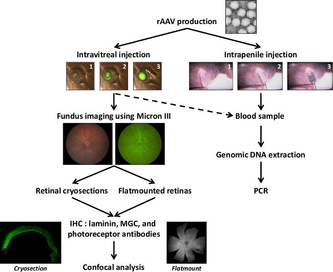
Figure 1: Schematic representation of the general protocol. After AAV production, particles are injected into the vitreous or into the penile vein to anesthetize adult mice. Fundus images with a Micron III camera are performed to follow the ...
Access restricted. Please log in or start a trial to view this content.
Materials
| Name | Company | Catalog Number | Comments |
|---|---|---|---|
| Superfrost Plus Adhesion Slides | Thermo Scientific | 10143352 | Slides |
| Anti-laminin | Sigma | L9393 | Antibody |
| Anti-rhodopsin clone 4D2 | Millipore | MABN15 | Antibody |
| Anti-glutamine synthetase clone GS-6 | Millipore | MAB302 | Antibody |
| Anti-Glial Fibrillary Acidic Protein | Dako | 334 | Antibody |
| PNA Lectin | Invitrogen | L32459 | Probe |
| Alexa fluor conjugated secondary antibodies | Invitrogen | Antibody | |
| Fluorsave reagent | Calbiochem | 345789 | Mounting medium |
| Evans Blue dye | Sigma | E2129 | Dye |
| Formamide spectrophotometric | Sigma | 295876-2L | |
| Fluorescein | Sigma | F2456 | Dye |
| Micron III | Phoenix Research Labs | Microscopy system based on 3-CCD color camera, frame grabber, and off-the-shelf software enables researchers to image mouse retinas. | |
| Binocular magnifier SZ76 | ADVILAB | ADV-76B2 | Zoom 0.66 x 5 x LEDs with stand epi and dia / Retinas dissection |
| Spring scissors straight - 8.5 cm | Bionic France S.a.r.l | 15003-08 | Retinal dissection |
