This video demonstrates the purification of prototype foamy virus (PFV) intasomes from a mixed protein-DNA suspension using salt extraction and size exclusion chromatography, yielding active complexes for downstream analysis.
A subscription to JoVE is required to view this content. Sign in or start your free trial.
Method Article
Purification of Prototype Foamy Virus Intasomes by Size Exclusion Chromatography
123 views
⸱
August 31st, 2026
In This Article
Abstract
Protocol
1. Intasome Solubilization
- Prepare two wide bore pipette tips by removing 2-4 mm from the end of a 200 µL tip with a new razor or scalpel blade on a clean surface.
NOTE: The tips will be used in step 1.3 and 1.5. - Remove the dialysis tubing from the dialysis buffer. In order to prevent dilution of the intasome sample with a dialysis buffer that may remain at the end of the tubing near the clip, use a micropipette with a small pipette tip to remove any excess dialysis buffer. Remove the clip from one end of the dialysis tubing.
- Use a wide bore pipette tip from step 1.1 to transfer the sample including the precipitate inside the dialysis tubing to a 1.5-mL tube on ice.
NOTE: The total volume of the recovered material is typically ~140 µL. - The sample with precipitate is at 200 mM NaCl; increase the salt to a final concentration of 320 mM NaCl by adding the appropriate volume of a stock 5 M NaCl solution. For example, for a sample of 150 µL, add 3.9 µL 5 M NaCl and 1.1 µL ddH2O for a final volume of 155 µL. Transfer ice bucket with sample into a 4 ˚C cold room.
- Use a wide bore pipette tip from step 1.1 to resuspend and solubilize the precipitate by pipetting. Repeat every 20 min for at least 1 h. Observe that most of the precipitate should go into solution.
NOTE: Pipetting to resuspend the precipitate can be stopped when it is apparent that the precipitate is no longer noticeably reduced between time points. When viralDNA (vDNA) is not labeled or is internally labeled, the precipitate is reduced by ~90% based on visual inspection. In the case of Cy5 end labeled vDNA, the precipitate is reduced by only ~20%; most of the precipitate with fluorophore end labeled vDNA will not solubilize. The amount of precipitate that is effectively solubilized is variable and should be empirically determined.
2. Intasome Purification
- Prepare 250 mL of size exclusion chromatography (SEC) running buffer (20 mM Bis-tris propane, pH 7.5, 320 mM NaCl, 10% glycerol). Sterile filter the buffer with a 0.2-µm filter unit and store at 4 ˚C.
NOTE: All purification steps are performed in a 4 ˚C cold room. - Equilibrate a cross-linked agarose SEC column (diameter = 10 mm; length = 300 mm; bed volume = 24 mL; sample volume = 25 - 500 µL; maximum pressure = 1.5 MPa; exclusion limit = 4 x 10⁷ Da; separates molecular weights between 5 and 5,000 kDa, see the Table of Materials) with SEC running buffer at a flow rate of 0.4 mL/min.
NOTE: This purification may be adapted to different size exclusion columns if they are able to effectively separate 300 kDa from 44 kDa, such as Superose 12 10/300 GL or Superose 6 Increase. Superose 12 10/300 GL has lower resolution, leading to more overlap of peaks. Conversely, Superose 6 Increase has higher resolution and offers better separation. - Centrifuge the intasome sample in a microfuge at 14,000 x g for 10 min at 4 ˚C to pellet any remaining precipitate. Carefully remove the supernatant. Load the supernatant to a 200 µL injection loop (tubing that can hold ~200 μL). Apply the sample to the SEC column.
NOTE: Smaller load volumes allow greater resolution by SEC. - Elute with 25 mL SEC running buffer and collect 95 fractions of 270 µL. Observe that the SEC chromatogram displays three A280/A260 peaks (Figure 1).
NOTE: The first should be an aggregate (~9.0 - 11.5 mL elution), followed by the PFV tetramer intasome peak (~12.0 - 14.75 mL elution) and PFV IN monomer with free vDNA peak (~15.0 - 18.0 mL).
Access restricted. Please log in or start a trial to view this content.
Results
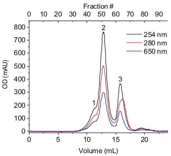
Figure 1: Size exclusion chromatogram. A PFV intasome assembly was separated with an agarose SEC column. This column allows for the separation of the aggregate (1), PFV intasome consisting of a tetramer of PFV IN and two vDNA (2), and monomeri...
Access restricted. Please log in or start a trial to view this content.
Materials
| Name | Company | Catalog Number | Comments |
|---|---|---|---|
| DNA Oligomers | IDT | N/A | Custom DNA Oligos |
| NaCl | P212121 | RP-S23020 | |
| BIS-TRIS propane,>=99.0% (titration) | Sigma-Aldrich | B6755-500G | |
| Glycerol | Thermo Fisher Scientific | G37-20 | |
| Gel-loading tips, 1 - 200 μL | Corning | CLS4853-400EA | |
| Razor blade; Single-edged; 100/Pk.; Pack of 100 | Fisher Scientific | 12-640 | |
| Dialysis tubing clips | Spectrum Labs | 132734 | |
| 6-8 kDa 10 mm Dialysis Tubing | Spectrum Medical | 132645 | |
| Superose 6 10/300 GL | GE Healthcare Life Sciences | 17517201 |
