This video demonstrates the isolation of protein–viral DNA complexes from cell nuclei using click chemistry and magnetic bead purification. The protocol involves labeling viral genomes with alkyne-modified nucleotides, biotinylation via a click reaction, and binding to streptavidin-coated magnetic beads. Following washing and heat elution, the purified complexes are collected, flash-frozen, and stored for downstream analysis.
A subscription to JoVE is required to view this content. Sign in or start your free trial.
Method Article
Isolation of Proteins from Viral DNA-protein Complexes
95 views
⸱
August 31st, 2026
In This Article
Abstract
Protocol
1. Purification of vDNA and Associated Proteins
NOTE: All buffers and reagents should be chilled on ice before use, and all steps should be carried out on ice unless otherwise indicated.
- Harvest nuclei.NOTE: Before isolation of nuclei, formaldehyde can be used to crosslink proteins to DNA. More stringent wash conditions can then be used during DNA purification on streptavidin-coated beads.
- Replace growth medium with 20 mL nuclei extraction buffer (NEB) and incubate for 20 min at 4 °C with occasional rocking. Nuclei will become visible in the microscope.
- Scrape nuclei from the plate using a cell scraper and transfer to a 50 mL conical tube. Centrifuge for 10 min at 2,500 x g at 4 °C to pellet nuclei, discard the supernatant.
NOTE: Trypan blue staining can be used to verify the isolation of nuclei from cells. - Gently dislodge the nuclear pellet in 10 mL phosphate-buffered saline (PBS), transfer to a 15-mL conical tube, and pellet by centrifuging for 10 min at 2,500 x g at 4 °C. Completely remove PBS.
NOTE: Nuclei can be frozen, and the protocol can be paused at this point. To do so, gently resuspend the nuclear pellet in a 500 µL freezing buffer and incubate the tube in an ethanol/dry ice bath. Store frozen nuclei at -80 °C. Before use, thaw nuclei at room temperature, quickly transfer to ice, add 10 mL PBS, rock gently to mix, pellet by centrifugation for 10 min at 2,500 x g at 4 °C, and completely remove PBS. Freezing nuclei may result in reduced yield.
- Covalently conjugate biotin-azide to 5-ethynyl-2′-deoxycytidine (EdC)-labeled viral DNA or vDNA.
- Resuspend the nuclear pellet in 10 mL click reaction mix [see Table of Materials] by gently pipetting up and down 5 times with a 10 mL pipette.
NOTE: It is important that the reagents included in the click reaction mix are added in the indicated order. - Rotate for 1h at 4 °C. While rotating, prepare and chill Buffers B1, B2, and B3 [see Table of Materials].
- Pellet nuclei by centrifuging for 10 min at 2,500 x g at 4 °C. Completely remove the click reaction mix and wash the pellet by gently resuspending in 10 mL PBS and centrifuging for 10 min at 2,500 x g at 4 °C.
- Gently resuspend the nuclear pellet in 1 mL PBS and transfer to a 1.5 mL microfuge tube using a large-bore pipette tip. Pellet nuclei by centrifuging for 10 min at 2,500 x g at 4 °C. Completely remove PBS and flash freeze in liquid nitrogen.
NOTE: The protocol can be paused at this point, and frozen nuclei can be stored at -80 °C. Freeze-thaw helps with subsequent lysis, so it is recommended that nuclei be flash-frozen even when proceeding to Step 1.3 on the same day.
- Resuspend the nuclear pellet in 10 mL click reaction mix [see Table of Materials] by gently pipetting up and down 5 times with a 10 mL pipette.
- Lyse nuclei and fragment DNA.
- Thaw nuclei on ice and resuspend in 500 µL Buffer B1 by pipetting up and down. Incubate on ice for 45 min.
- Sonicate samples 6 times for 30 s each at 40% amplitude using a 3 mm microtip probe. Place samples on ice for 30 s between pulses. After sonication, samples should appear clear, not cloudy.
NOTE: Sonication conditions should be optimized for individual sonicators. - Pellet cell debris by centrifuging at 14,000 x g for 10 min at 4 °C. The pellet size should decrease substantially. Filter the supernatant through a 100 µM cell strainer and retain the flow through.
- Add 500 µL Buffer B2 to the filtered supernatant. 900 µL of this sample will be used in Step 1.4.2.
- Take an aliquot of each condition for DNA isolation (50 µL or 1/20th volume) and add 50 µL 2x sodium dodecyl sulfate (SDS)-bicarb solution [see Table of Materials]. Proceed to the DNA isolation protocol.
- Take an aliquot of each condition for input protein (50 µL or 1/20th volume) and add 50 µL 2x Laemmli sample buffer, freeze in liquid nitrogen, and store at -80 °C.
- Bind biotinylated DNA to streptavidin-coated beads.
- Prepare Streptavidin T1 magnetic beads by transferring 300 µL of bead slurry to a 1.5 mL microfuge tube. Prepare one tube of beads per sample. Wash beads 3x with 1 mL Buffer B2 by vortexing to resuspend, applying a magnet to separate the beads, and aspirating the wash buffer.
NOTE: Optimized binding conditions result in maximized yield and minimal background binding. It was experimentally determined that Streptavidin T1 magnetic beads have a significantly greater binding capacity than agarose beads, as well as other Streptavidin beads, and less background binding than Streptavidin C1 beads (Figure 1A). - Add 900 µL of the sample from Step 1.3.4. to the washed beads and rotate overnight at 4 °C.
Note: Do not vortex beads once the sample is added to them.
NOTE: If significant amounts of DNA or protein are isolated in the unlabeled negative control, this step can be reduced from overnight to 4 h to reduce background binding.
- Prepare Streptavidin T1 magnetic beads by transferring 300 µL of bead slurry to a 1.5 mL microfuge tube. Prepare one tube of beads per sample. Wash beads 3x with 1 mL Buffer B2 by vortexing to resuspend, applying a magnet to separate the beads, and aspirating the wash buffer.
- Wash beads and elute vDNA and associated proteins.
NOTE: It is recommended to use filter tips for the remaining steps.- Place samples into a magnetic microfuge tube rack, remove supernatant, gently resuspend in 1 mL Buffer B2, and rotate at 4 °C for 5 min.
- Repeat Step 1.5.1 three times.
- Place the samples into a magnetic microfuge tube rack, remove supernatant, gently resuspend in 1 mL Buffer B3, and rotate at 4 °C for 5 min.
- Transfer 100 µL bead mixture (1/10th volume) to a new tube for bound DNA isolation. Apply this tube to the magnet, remove supernatant, resuspend beads in 100 µL 1x sodium dodecyl sulfate or SDS-bicarb solution, and proceed to the DNA isolation protocol.
- Apply the tube containing the remaining 900 µL bead mixture from step 1.5.3. to the magnet, remove supernatant, and resuspend beads in 50 µL 2x Laemmli sample buffer to elute protein and DNA-protein complexes.
- Boil samples at 95 °C for 15 min, vortex, quickly spin in the microfuge, and apply the magnet. Transfer eluate to a new tube, flash freeze, and store at -80 °C.
NOTE: Use the caps lock to ensure that the tubes do not pop open while boiling.
NOTE: The protocol can be paused at this point, and samples can be stored at -80 °C for several weeks. - Analyze protein samples by Coomassie Blue staining, western blotting, or mass spectrometry by standard procedures. Representative results are shown in Figures 1 and 2.
NOTE: To determine if protein yield is sufficient for mass spectrometry, 7.5 µL of each sample is used for analysis by Coomassie Blue staining and 7.5 µL for western blotting of a representative viral genome-associated protein (ICP4, ICP8, or UL42). The same volume of lysates from Step 1.3.6. are run alongside to control for input protein levels. The remaining sample (35 µL) is then analyzed by mass spectrometry.
Access restricted. Please log in or start a trial to view this content.
Results
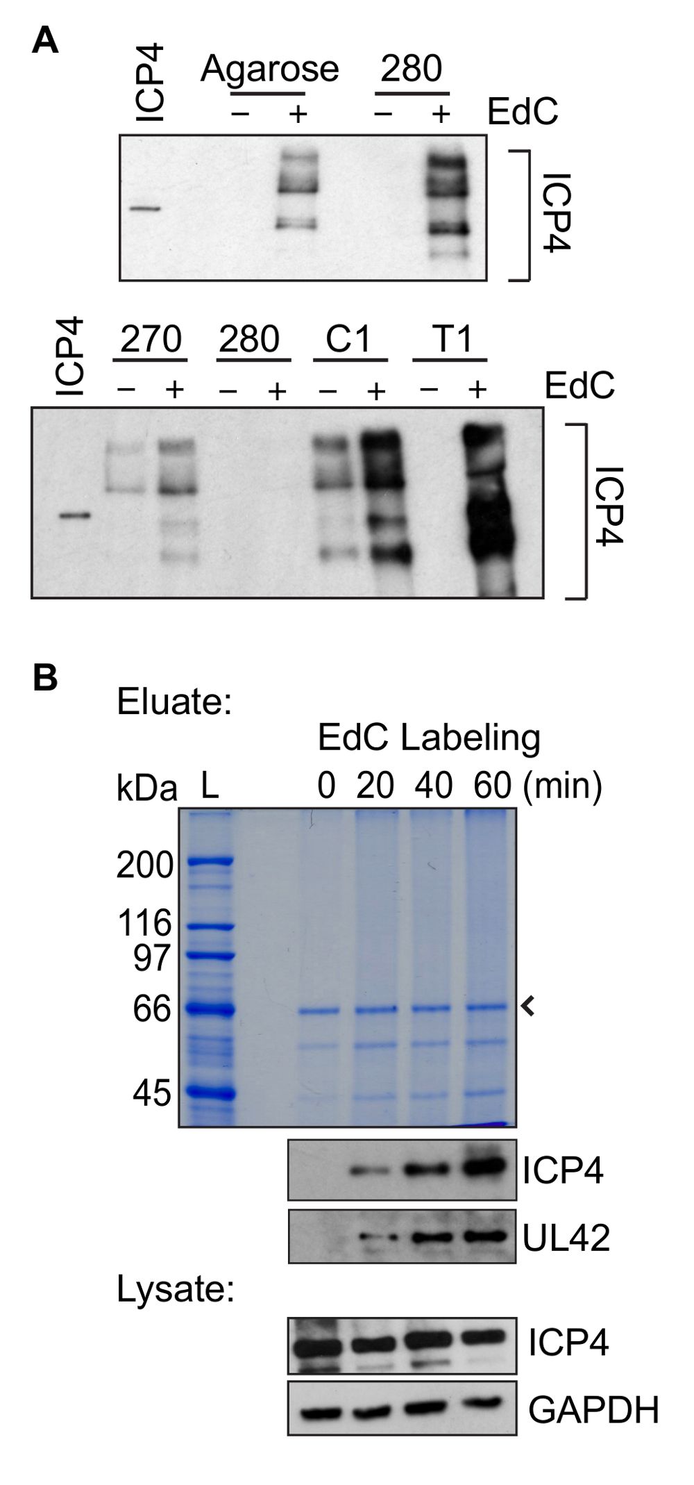
Figure 1: Representative protein purification results. (A) Comparison of protein yield when purification steps were carried out using several different types of streptavidin-coated beads. Infection was carried out in the p...
Access restricted. Please log in or start a trial to view this content.
Materials
| Name | Company | Catalog Number | Comments |
|---|---|---|---|
| MRC-5 cells | ATCC | CCL-171 | |
| Fetal Bovine Serum (FBS) | Gibco | 26140-179 | |
| Dulbecco's Modified Eagle Medium (DMEM) | Gibco | 12800-082 | Substituted with 10% FBS, 2 mM L-glutamine, 12 mM (for growth in flasks) or 30 mM (for growth in dishes) sodium-bicarbinate |
| 600 cm² tissue culture dish | Thermo Fisher Scientific | 166508 | |
| Tris Buffered Saline (TBS), pH 7.4 | 137 mM NaCl, 5 mM KCl, 491 mM MgCl, 680 mM CaCl, 25.1 mM Tricine | ||
| HSV-1 stock | Stocks with titers greater than 1x10⁹ PFU/mL work best | ||
| Sephadex G-25 column (PD-10 Desalting Column) | GE Healthcare | 17085101 | |
| Dimethyl sulfoxide (DMSO) | Fisher Scientific | D128-1 | |
| 5´-Ethynyl-2´-deoxycytidine (EdC) | Sigma-Aldrich | T511307 | Dissolve in DMSO to prepare 40 mM stock, aliquot, and store at -20 °C |
| 2´-deoxycytidine (deoxyC) | Sigma-Aldrich | D3897 | Dissolve in water to prepare 40 mM stock, aliquot, and store at -20 °C |
| Nuclear Extraction Buffer (NEB) | Prepare fresh (20 mM Hepes pH 7.2, 50 mM NaCl, 3 mM MgCl₂, 300 mM Sucrose, 0.5% Igepal) | ||
| Cell scraper | Bellco glass | 7731-22000 | Autoclave before use |
| Trypan blue solution | Sigma-Aldrich | T8154 | |
| PBS, pH 7.2 (10x) | 1.37 M NaCl, 27 mM KCl, 100 mM Na₂HPO₄, 18 mM KH₂PO₄ (dilute to 1x in sterile water before use) | ||
| Copper (II) sulfate pentahydrate (CuSO₄·5H₂O) | Fisher Scientific | C489 | Prepare 100 mM stock and store at 4 °C for up to 1 month |
| (+) Sodium L-ascorbate | Sigma-Aldrich | A4034 | Freshly prepare 100 mM stock and store on ice until use |
| Biotin azide | Invitrogen | B10184 | Prepare 10 mM stock in DMSO, aliquot, and store at -20 °C for up to 1 year |
| Click Reaction Mix | Prepare immediately before use by adding reagents in the indicated order (10 mL: 8.8 mL 1x PBS, 200 mL 100 mM CuSO₄, 25mL 10 mM Biotin Azide, 1 mL 100 mM sodium ascorbate) | ||
| Complete Protease Inhibitor Cocktail | Roche | 11697498001 | Dissolve in 1 mL water to prepare 50x stock, can store at 4 °C for up to 1 week, or directly add 1 pill to 50 mL buffer |
| Freezing buffer | Prepare fresh (7 mL 100% glycerol, 3 mL NEB, 200 μL 50x protease inhibitor) | ||
| Buffer B1 | Prepare fresh (25 mM NaCl, 2 mM EDTA, 50 mM Tris-HCl pH 8, 1% Igepal, 1x protease inhibitor) | ||
| Buffer B2 | Prepare fresh (150 mM NaCl, 2 mM EDTA, 50 mM Tris-HCl pH 8, 0.5% Igepal, 1x protease inhibitor) | ||
| Buffer B3 | Prepare fresh (150 mM NaCl, 2 mM EDTA, 50 mM Tris-HCl pH 8, 1x protease inhibitor) | ||
| Vibra Cell Ultra Sonic Processer equipped with a 3 mm microtip probe | Sonics | VCX 130 | |
| Cell strainer | Falcon | 352360 | |
| Dynabeads MyOne Streptavidin T1 | Life Technologies | 65601 | |
| DynaMag-2 Magnet | Life Technologies | 12321D | |
| Mini-Tube Rotator | Fisher Scientific | 260750F | |
| 2x Laemmli sample buffer | Mix 400 mg of SDS, 2 mL 100% glycerol, 1.25 mL of 1 M Tris (pH 6.8), and 10 mg of bromophenol blue in 8 mL water. Store at 4 °C for up to 6 months. Before use add 1 M DTT to a final concentration of 200 mM. | ||
| Cap locks for 1.5 mL tube | Fisher Scientific | NC9679153 | |
| Standard western blotting reagents | |||
| NOVEX Colloidal Blue Staining Kit | Invitrogen | LC6025 | |
| 12-well tissue culture dish | Corning | 3513 | |
| Coverslips | Fisher Scientific | 12-545-100 | Autoclave before use |
| Microscope slides | Fisher Scientific | 12-552-5 | |
| 16% paraformaldehyde | Electron Microscopy Sciences | 15710 | Dilute to 3.7% in 1x PBS before use |
| Bovine serum albumin (BSA) | Fisher Scientific | BP1605 | Prepare 3% in 1x PBS |
| Permeabilization buffer (0.5% Triton-X 100) | Sigma-Aldrich | T8787 | Prepare 0.5% Triton-X 100 in 1x PBS |
| Click reaction cocktail - Click-iT EdU Alexa Fluor 488 Imaging Kit | Molecular Probes | C10337 | Prepare according to manufactorer's protocol |
| 2x SDS-bicarb solution | 2% SDS, 200 mM NaHCO₃ | ||
| 10x Tris EDTA (TE), pH 8.0 | 100 mM Tris, 10 mM EDTA (dilute to 1x before use) | ||
| Microcentrifuge for 1.5 mL tubes | |||
| Tabletop centrifuge for 15 and 50 mL tubes | |||
| Cell culture incubator | |||
| Biosafety cabinet | |||
| Heat blocks | 65 °C and 95 °C |
