This video demonstrates the purification of high-capacity adenoviral vectors using cesium chloride gradient ultracentrifugation, enabling the separation of genome-containing virions for applications in gene delivery, vector characterization, and preclinical therapeutic studies
A subscription to JoVE is required to view this content. Sign in or start your free trial.
Method Article
Cesium Chloride-Based Purification of Genome-Containing Adenoviral Vectors
103 views
⸱
August 31st, 2026
In This Article
Abstract
Protocol
- Purification and Dialysis of HCAdV (High-capacity adenoviral vectors)
- To prepare viral lysate for cesium chloride (CsCl) gradients, freeze the cell-virus suspension in liquid nitrogen and thaw in a water bath at 37 °C 4x.
- Centrifuge the viral lysate at 500 x g for 8 min at RT (room temperature) and collect the supernatant containing the HCAdV (high-capacity adenoviral vectors) .
- For ultracentrifugation, prepare CsCl solutions as follows. Weigh 37.5 g (for 1.5 g/cm3), 33.5 g (for 1.35 g/cm3), and 31.25 g (for 1.25 g/cm3) of CsCl powder, respectively, and fill up with dH2O to 25 ml.
- Stir until the solution becomes clear and sterile filter the solutions. Finally remove 1 ml of each solution and weigh it on a fine scale to check whether the density is correct. 1 ml should weigh 1.5 g, 1.35, and 1.25 g respectively.
- If the density is too high, adjust it by stepwise addition of small volumes (µl) of sterile dH2O. After addition of dH2O measure the density again. If necessary add more dH2O.
- Repeat the procedure until the density is correct. If the density is too low, adjust it by adding a small amount of CsCl powder. Sterile filter it again and check the density. If the density is too high, adjust it by adding dH2O as described before. If the density is still too low add more CsCl, sterile filter it again and check the density. Repeat the procedure until the density is correct.
- Prepare CsCl step gradients in 6 clear ultracentrifuge tubes. Carefully and slowly pipette CsCl solutions into the tubes using the following order: 0.5 ml of 1.5 g/cm3 CsCl solution, 3 ml of 1.35 g/cm3 CsCl solution, and 3.5 ml of 1.25 g/cm3 CsCl solution (Figure 1C).
- Overlay ~4.5 ml of cleared vector supernatant from step 1.2 on top of the 1.25 g/cm3 CsCl layer. Centrifuge the gradients in an ultracentrifuge using a swing-out rotor (SW-41) at 12 °C for at least 2 hr at 226,000 x g (35,000 rpm) with slow acceleration and deceleration to separate HCAdV-genome containing viral particles from empty particles and cell debris.
NOTE: Under optimal conditions a diffuse band of cell debris formed on top of the tubes. Below, two white bands can be observed. The upper band contains empty particles whereas the lower band equals the HCAdV (Figures 1C and 2A). - Carefully remove the layers of cell debris and empty particles and collect 1 ml of the lower bands from each tube and transfer virus with a clean pipette tip into a sterile 50 ml tube. Add up to 24 ml of 1.35 g/cm³ CsCl solution to the collected virus particles and mix carefully.
- Fill centrifuge tubes with 1.35 g/cm3 CsCl-virus solution to the top and centrifuge O/N (overnight) (18-20 h) at 226,000 x g (35,000 rpm) at 12 °C in an ultracentrifuge using a swing-out rotor (SW-41) with slow acceleration and deceleration.
- Collect the HCAdV present in the prominent lower band. As a potential upper band contains empty particles, remove upper layers from the top using a pipette (see Figures 1C and 2B). Then, take away the lower band using a pipette.
- Dialyze the collected virus particles for buffer exchange.
- Cut off a strip of dialysis tubing (MWCO: 50,000) of about 8 cm in length, wash it with sterile dH2O for 3x. Then close one side of the dialysis tubing with a plastic clamp and transfer the collected virus from step 5.8 into tubing using a 1-ml pipette. Avoid air bubbles within the tubing and close it on the other side with a plastic clamp dialysis closure.
- Dialyze in 1-L of dialysis buffer (10 mM Tris-HCl (pH 7.5), 10% glycerol, and 1 mM MgCl2 (magnesium chloride) in deionized H2O) for 2 hr at 4 °C with slow stirring. Exchange dialysis buffer with 2 L of dialysis buffer and dialyze O/N at 4 °C with slow stirring. Alternatively, use a sucrose buffer (140 mM NaCl (sodium chloride), 5 mM Na2HPO4.2H2O (di-sodium hydrogen phosphate, 1.5 mM KH2PO4 (potassium dihydrogen phosphate), and 730 mM sucrose, pH 7.8).
- Collect dialyzed virus particles from step 1.9.2. using a 1 ml pipette. Prepare multiple aliquots of desired small volumes (25-100 µl) and store purified virus at -80 °C.
Access restricted. Please log in or start a trial to view this content.
Results
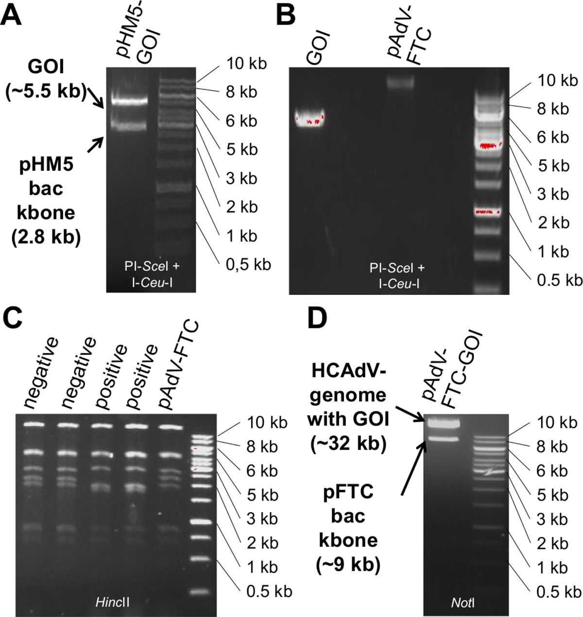
Figure 1. Representative results for the cloning procedure and release of the HCAdV (High-capacity adenoviral vectors) genome from pAdVFTC. (A) Release of gene of interest (GOI)-expression cassette from pHM5 by PI-SceI and I-Ceu<...
Access restricted. Please log in or start a trial to view this content.
Materials
| Name | Company | Catalog Number | Comments |
|---|---|---|---|
| Ultra Clear Ultracentrifuge tubes | Beckmann Coulter | 344059 | density gradient centrifugation |
| Ultracentrifuge | Beckmann Coulter | density gradient centrifugation | |
| SW-41 rotor | Beckmann Coulter | density gradient centrifugation | |
| Spectrum Laboratories Spectrapor Membrane | VWR | 132129 | dialysis tubing |
| Ready-to-use dialysis cassettes | Thermo | 66383 | dialysis |
| Cesium chloride | Carl Roth | 8627.1 | density gradient centrifugation |
| LB medium | Carl Roth | X968.3 | bacterial growth medium |
| Tris-HCl 99% | Carl Roth | 9090.3 | dialysis buffer/ lysis buffer |
| Glycerol 99.5% | Carl Roth | 3783.1 | dialysis buffer |
| MgCl₂ 98.5% | Carl Roth | KK36.2 | dialysis buffer |
| NaCl | Carl Roth | 3957.1 | optional dialysis buffer |
| KH₂PO₄ | Carl Roth | 3904.2 | optional dialysis buffer |
| Sucrose | Carl Roth | 9286.1 | optional dialysis buffer |
| Na₂HPO₄·2H₂O | Carl Roth | 4984.2 | optional dialysis buffer |
| 1.5 ml tubes | sarstedt | 72,730,005 | storage of virus preparations at -80 °C |
