The video demonstrates purification of recombinant adeno-associated virus (AAV) vectors from crude host lysate using a heparin-based affinity matrix. Viral particles bind to immobilized heparin, while impurities are washed away. A high-salt buffer releases intact viruses, which is collected in enriched fractions. These fractions are concentrated by membrane centrifugation and rinsed to recover residual particles, producing purified, high-titer AAV vectors.
A subscription to JoVE is required to view this content. Sign in or start your free trial.
Method Article
Affinity Chromatography-Based Purification of Adeno-Associated Virus Vectors
125 views
⸱
August 31st, 2026
In This Article
Abstract
Protocol
1. Double plasmid transfection of HEK293 cells in cell stacks
- Thaw a cryo-vial of Human Embryonic Kidney 293 (HEK293) cells in a bead bath set at 37 °C.
NOTE: Pre-warm complete Dulbecco’s Modified Eagle Medium (DMEM) to 37 °C while cells thaw to prevent cold shock during plating. Ensure the cells have a low passage number, ideally less than 20, to ensure optimal growth and transfection efficiency. Ensure that the cells are certified to be mycoplasma-free. - Transfer the contents of the cryo-vial dropwise into a 15 mL conical tube containing 10 mL of pre-warmed complete DMEM and centrifuge the cells at 500 × g for 5 min.
- Aspirate the media, and then resuspend the HEK293 cells in 20 mL of pre-warmed complete DMEM. Seed the cells in a 15 cm plate and incubate at 37 °C, with 5% CO2.
- Split the cells from one 15 cm plate into three for seeding in the cell culture chamber.
- Once cells are 80% confluent, aspirate the media and gently wash the plate with 3 mL of phosphate-buffered saline (PBS) to not disrupt the monolayer. Then, aspirate PBS and add 3 mL of trypsin.
- Incubate for 2 min at 37 °C until the cells lift from the plate, and then neutralize trypsin by adding 7 mL of complete DMEM to the plate.
- Collect all the media and cells into a 15 mL tube and pellet the cells by centrifuging at 500 × g for 5 min.
- Aspirate the supernatant from the 15 mL tube and resuspend the cell pellet in 3 mL of complete DMEM. Add 1 mL to each 15 cm plate containing 20 mL of complete DMEM; gently rock the plates to distribute the cells evenly, and incubate at 37 °C, with 5% CO2.
- Once the cells are 80% confluent, repeat steps 1.4.1 and 1.4.2. Collect the supernatant in 50 mL conical tubes, and gently invert the tube to ensure the cells are homogeneous.
- Determine the cell density by mixing 10 µL of the samples of cells with 10 µL of trypan blue and adding the mixture to a cell counting slide for analysis in the cell counter.
- Mix 1 L of pre-warmed complete DMEM with the needed cell suspension to seed the cell culture chamber (surface area of 6360 cm2) with 1 × 10⁴ cells/cm2. Pour the cell mixture into the cell culture chamber and gently rotate to evenly distribute the cells across each monolayer (Figure 1) and incubate at 37 °C, with 5% CO2.
- In addition to the cell culture chamber, plate a 15 cm plate with 1 × 10⁴ cells/cm2 as a reference for confluency.
- Following ~65-h incubation, check the reference plate for confluency-ideally ~80%-90% confluent.
NOTE: Pre-warm complete DMEM for adding to the cell culture chamber at 37 °C. - Prepare polyethyleneimine (PEI)/DNA mixture at a concentration ratio of 3:1 (w/w).
- Prepare the DNA mixture in a 50 mL conical tube by adding 475 µg of pTrangene and 1425 µg of pHelper to 40 mL of reduced-serum medium to create a 3:1 ratio of pHelper:pTrangene.
NOTE: The PEI/DNA mixture calculator can be found using Table 1.
- Prepare the DNA mixture in a 50 mL conical tube by adding 475 µg of pTrangene and 1425 µg of pHelper to 40 mL of reduced-serum medium to create a 3:1 ratio of pHelper:pTrangene.
| Transfection of Cell Culture Chamber | |||||
| Protocol | |||||
| 1. Allow DNA, OptiMEM and PEI to warm to room temperature prior to transfection | |||||
| 2. Input the number of cell stacks to be transfected (no overage required) | |||||
| 3. Ensure the DNA concentrations are correct as this will change the volumes required | |||||
| Number of Cell Culture Chambers | 1 | ||||
| Concentration of transgene plasmid | 1 | mg/mL | |||
| Concentration of pDGM6.2FF plasmid | 1 | mg/mL | |||
| Amount Per cell culture chambers | Transfection Mastermix | ||||
| OptiMEM | 48.1 | mL | 48.1 | mL | |
| 1:3 ratio pTransgene:pHelper | pTransgene | 475 | µg | 475 | µL |
| pHelper | 1425 | µg | 1425 | µL | |
| 4. Add required volumes of OptiMEM and plasmid DNA to a 50 mL conical tube | |||||
| 5. Invert 10 times to mix | |||||
| 3:1 PEI:DNA ratio | PEI Max | 5.7 | mL | 5.7 | mL |
| 6. Add the required amount of PEI to the 50 mL tube containing the OptiMEM and plasmid DNA | |||||
| 7. Immediately close the 50 mL tube and vortex and invert 3–5 times to mix. | |||||
| 8. Set a timer for 10 min for the PEI complexes to incubate | |||||
| 9. Move cell stacks to be transfected into the Biological Safety Cabinet (BSC) | |||||
| 10. After 10 min, pour the transfection mix into one orange port. | |||||
| 11. Gently mix liquid throughout the cell stack | |||||
| 12. Return the transfected cells stack to the incubator ensuring equal volume on each layer | |||||
| 13. Harvest cell culture chamber 72 h later | |||||
Table 1: Transfection calculator for cell culture chamber. Interactive worksheet to determine correct concentration of pTransgene, pHelper, and PEI for transfection of cell culture chamber.
2. Add 5.7 mL of PEI (1 g/L) to the reduced-serum medium and the DNA mixture dropwise. Then, vortex briefly and incubate for 10 min at room temperature.
NOTE: As PEI/DNA incubates at room temperature, it will become slightly cloudy.
- After 8 min of PEI/DNA incubation, remove the media from the cell culture chamber.
NOTE: Ensure to loosen both orange caps to maintain a smooth flow of media to avoid dislodging of cells. - Add PEI/DNA to 1 L of pre-warmed complete DMEM, and slowly pour the mixture into the cell culture chamber port. Distribute the liquid evenly to all the rows (Figure 1) and incubate for 72 h at 37 °C, with 5% CO2.
2 Harvesting adeno-associated virus (AAV) and chemical lysis of the transfected HEK293 cells
- Shake the cell culture chamber vigorously to dislodge the cells until the media appears cloudy from dislodged cells and pour into four 500 mL centrifuge tubes.
- Centrifuge the tubes at 18,000 × g for 30 min at 4 °C to pellet the cells. Pour the clarified supernatant into 1 L polyethylene terephthalate copolyester (PETG) bottle.
NOTE: If one does not have access to a high-speed centrifuge, centrifuge at 12,000 × g for 40 min. The pelleted cells may not be solid at this speed and will slide as they pour out supernatant. - Resuspend the cell pellets in 500 mL centrifuge tubes with 50 mL of lysis buffer and incubate for 60 min at 37 °C.
- Centrifuge the tubes at 18,000 × g for 30 min, and then transfer the supernatant into the same 1 L PETG bottle. Discard the pelleted cell debris.
NOTE: Purify the clarified supernatant immediately and store at 4 °C for up to 72 h. For longer-term storage, store at -80 °C. Do not store at -20 °C.
3 AAV Vector purification using heparin affinity chromatography
- Remove the crude lysate from -80 °C and leave at 4 °C overnight to thaw. Once thawed, use a 0.22 µM filter to filter the crude lysate.
- To passivate the centrifugal concentrator, add 4 mL of filter pre-treatment buffer to a centrifugal concentrator for each heparin sepharose column being used. Passivate the centrifugal concentrator at room temperature for 2-8 h. Set up passivation immediately before the purification steps.
- Set up the tubing and the pump (Figure 2).
- Place the tubing in a peristaltic pump and run 20 mL of 1 M NaOH. Next, run 50 mL of molecular grade water, and then run 50 mL of basal DMEM.
- Attach 5 mL heparin sepharose column to tubing and run 25 mL of basal DMEM to remove the preservative.
- Run 0.2 µM of the filtered crude lysate through the column at a flow rate of 1-2 drops/s.
NOTE: Ensure not to introduce bubbles or allow the column to run dry, as this will compromise the column and prevent the elution of AAV. Discard the column if it runs dry and use a new column for the remainder of crude lysate.
- Load all of the crude lysate onto the heparin column and use the following solutions to wash the column.
- Wash using 50 mL of 1× Hank's Balanced Salt Solutions (HBSS) without Mg2+ and Ca2+.
- Wash using 15 mL of 0.5 % N-Lauroylsarcosine in HBSS without Mg2+ and Ca2+.
- Wash using 50 mL of HBSS without Mg2+ and Ca2+.
- Wash using 50 mL of HBSS with Mg2+ and Ca2+
- Wash using 50 mL of 200 mM NaCl/HBSS with Mg2+ and Ca2+.
- Elute 5 × 5 mL (25 mL total) with 300 mM of NaCl/HBSS with Mg2+ and Ca2+ and label the elutions as E1-E5 (each elution is of 5 mL).
- Concentrating the virus using a centrifugal concentrator
- Spin the centrifugal concentrator containing the pre-treatment buffer at 900 × g for 2 min. Discard the flow-through.
- Wash the centrifugal concentrator filter with 4 mL of HBSS with Mg2+ and Ca2+ and centrifuge at 1000 × g for 2 min; discard the flow-through.
- Add the elution E2 to the centrifugal concentrator. Spin at 1000 × g for 5 min and discard the flow-through.
- Finish adding E2 and then add E3 to the centrifugal concentrator and spin at 1000 × g for 5 min until the concentrated virus is approximately 1 mL.
NOTE: Avoid centrifuging the vector such that the volume is below the level of the filter. Do not concentrate E1, E4, or E5 in the centrifugal concentrator, as they contain very little to no vector and contain contaminants. - Remove the concentrated virus from the centrifugal concentrator using a p200 filtered tip and place it into a sterile 1.5 mL centrifuge tube.
- Rinse the centrifugal concentrator with 200 µL of HBSS with Mg2+ and Ca2+ to dislodge any remaining AAV from the filter. Pipette up and down vigorously multiple times (for ~30 s) to dislodge any virus adhered to the membrane and place in the 1.5 mL centrifuge tube with the remainder of the virus. Mix the tube well.
- Aliquot 5 µL for DNA extraction and store the purified vector at -80 °C.
- Wash the column using 25 mL of 2 M NaCl. Further, use 25 mL of 0.1% Triton X-100, pre-heated to 37 °C to wash the column. Next, wash the column using 50 mL of sterile dH2O, and then wash using 25 mL of 20% ethanol.
- Ensure the column membrane is fully saturated in 20% ethanol, as this is the storage solution. Seal the column with plugs provided and store at 4 °C.
- Store the tubing in 1 M NaOH.
NOTE: If cleaned properly, heparin sepharose columns can be re-used up to five times.
Access restricted. Please log in or start a trial to view this content.
Results
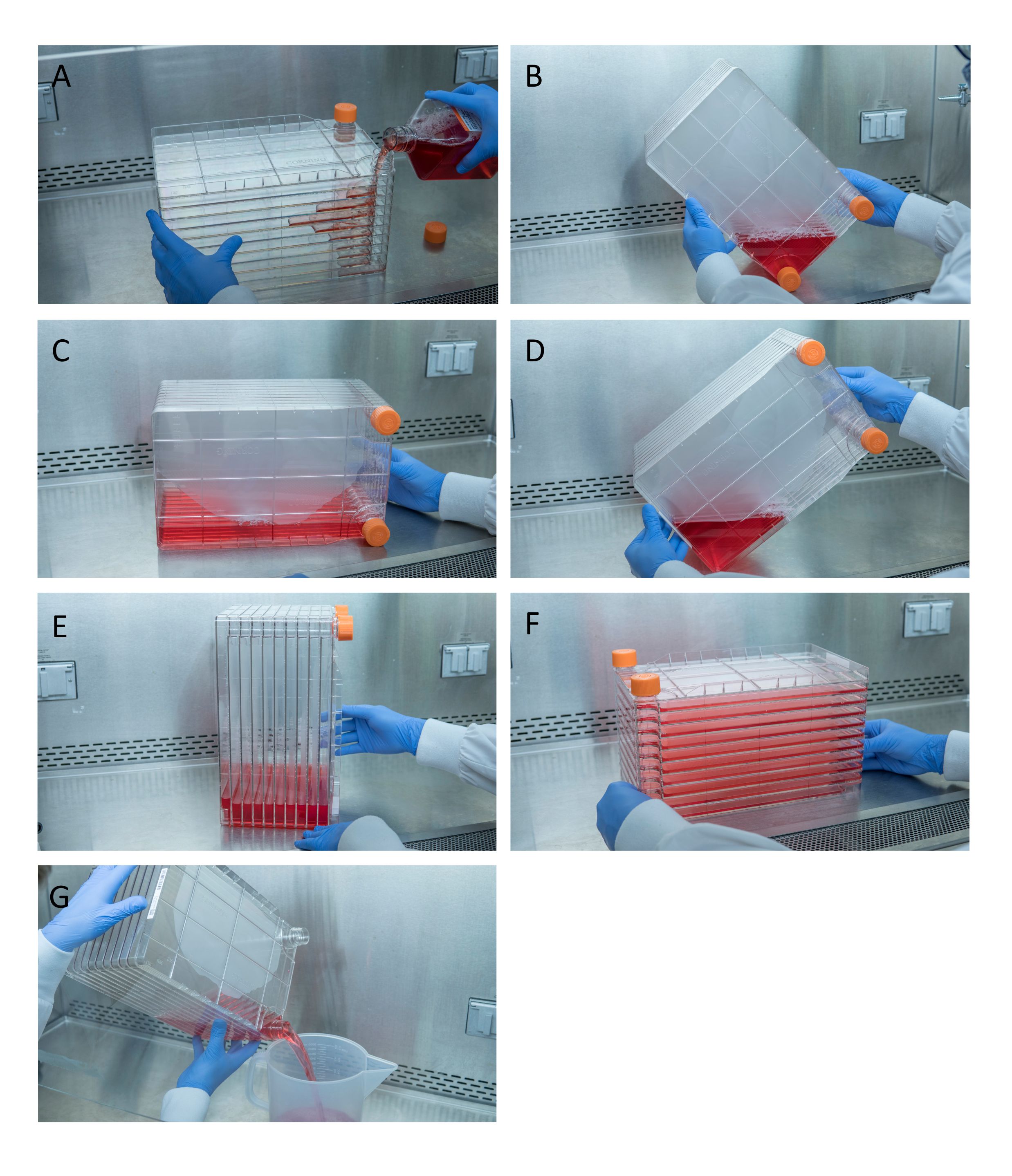
Figure 1: Maneuvering of cell stack for cell seeding and transfection. For seeding cell stack, start by removing one of the vent caps and pouring in 1 L of pre-warmed complete Dulbecco’s Modified Eagle Medium (DMEM) with needed quan...
Access restricted. Please log in or start a trial to view this content.
Materials
| Name | Company | Catalog Number | Comments |
|---|---|---|---|
| 0.22 μm Filter | Millipore Sigma | S2GPU05RE | |
| 0.25% Trypsin | Fisher Scientific | SM2001C | |
| 10 Chamber cellstack | Corning | 3271 | |
| 1L PETG bottle | Thermo Fisher Scientific | 2019-1000 | |
| Cell Culture Dishes | Greiner bio-one | 7000232 | 15 cm plates |
| Culture Conical Tube | Thermo Fisher Scientific | 339650 | 15 mL conical tube |
| Culture Conical Tube | Fisher Scientific | 14955240 | 50 mL conical tube |
| Dulbecco's Modified Eagle Medium (DMEM) with 1000 mg/L D-glucose, L-glutamine | Cytiva Life Sciences | SH30022.01 | |
| Ethanol | Greenfield | P016EA95 | Dilute ethyl alcohol(95% vol) to 20% for section 3.7.4 |
| Fetal Bovine Serum (FBS) | Thermo Fisher Scientific | SH30396.03 | |
| Hank's Balanced Salt Solutions (HBSS) with Mg²⁺ and Ca²⁺ | Thermo Fisher Scientific | SH302268.02 | |
| Hank's Balanced Salt Solutions (HBSS) without Mg²⁺ and Ca²⁺ | Thermo Fisher Scientific | SH30588.02 | |
| HEK293 cells | American Tissue Culture Collection | CRL-1573 | Upon receipt, thaw the cells and culture as described in manufacturer’s protocol. Once cells have been minimally passaged and are growing well, freeze a subfraction for future in aliquots and store in liquid nitrogen. Always use cells below passage number 30. Once cultured cells have been passaged more than 30 times, it is recommended to restart a culture from the stored aliquots |
| Heparin sulfate column | Cytiva Life Sciences | 17040703 | |
| Large Volume Centrifuge Tube Support Cushion | Corning | CLS431124 | Support cushion must be used with large volume centrifuge tubes uless the centrifuge rotor has the approriate V-bottom cushions |
| Large Volume Centrifuge Tubes | Corning | CLS431123-6EA | 500 mL centrifuge tubes |
| Microcentrifuge tube | Fisher Scientific | 05-408-129 | 1.5 mL microcentrifuge tube, sterilize prior to use |
| Molecular Grade Water | Cytiva Life Sciences | SH30538.03 | |
| N-Lauroylsarcosine sodium salt | Sigma Aldrich | L5125 | CAUTION. Wear gloves |
| NaCl | Thermo Fisher Scientific | BP35810 | |
| Optimem, reduced serum medium | Thermo Fisher Scientific | 31985070 | |
| Pasteur pipets | Fisher Scientific | 13-678-20D | Sterilize prior to use |
| Phosphate-buffered saline (10x) | Thermo Fisher Scientific | 70011044 | Dilute to 1x for use on cells |
| Penicillin-Streptomycin Solution | Cytiva Life Sciences | SV30010 | |
| pHelper plasmid | De novo design or obtained from plasmid repository | NA | |
| Pipet basin | Thermo Fisher Scientific | 13-681-502 | Purchase sterile pipet basins |
| Polyethylene glycol tert-octylphenyl ether (Triton X-100) | Thermo Fisher Scientific | 9002-93-1 | CAUTION. Wear gloves |
| Polyethylenimine (PEI) | Polyscience | 24765-1 | Follow manufacturer’s instructions to produce a 1L solution. 0.22μm filter and store at 4°C |
| pTrangene plasmid | De novo design or obtained from plasmid repository | NA | Must contain SV40 polyA in genome to be compatible with AAV titration in section 5.0 |
| Pump tubing | Cole-Parmer | RK-96440-14 | Optimize length of tubing and containment of virus in fractions E1-E5 |
| Sodium hydroxide (NaOH) | Thermo Fisher Scientific | SS266-4 | |
| Trypan blue | Bio-Rad | 1450021 |
