1. Construction of a concentrated lentivirus packaging system for transcription factors
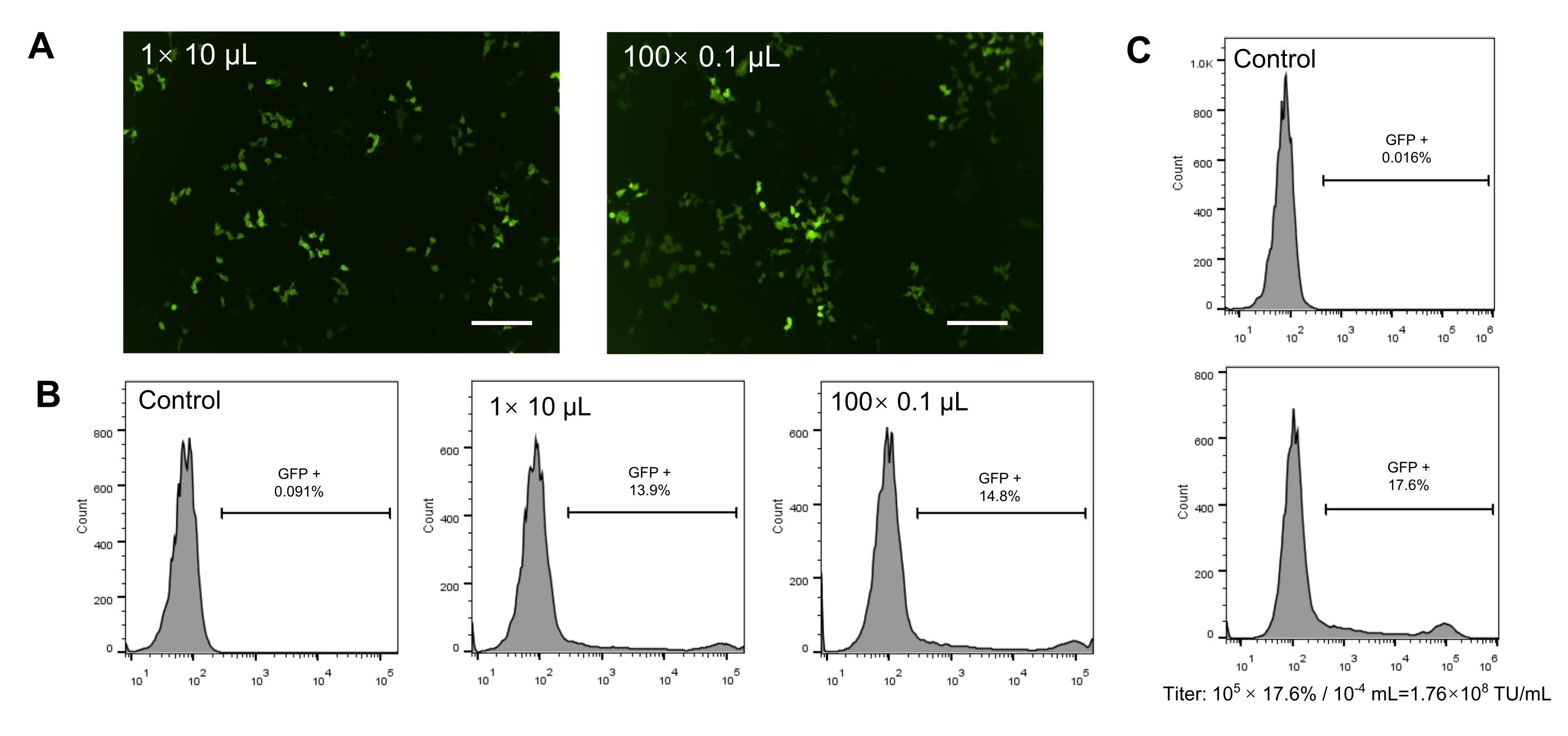
- Production of the concentrated virus (Figure 1A, B)
- Plate 1.5 × 10⁶ HEK (human embryonic kidney)-293T cells into a 60 mm dish and culture these cells with normal medium (see Table 1) at 37 °C in a humidified incubator with 5% CO2.
NOTE: If the virus needs to be packaged in batches, a 100 mm cell culture dish can be used (for details, see Table 2). - After 24 h, ensure that the HEK-293T cells have reached 80-90% confluence on the day of transduction, and replace the medium with 3.5 mL of DMEM (Dulbecco’s modified eagle medium) (150 µL of DMEM per 1 cm2). Leave the cells at 37 °C in a humidified incubator with 5% CO2 for 2 h.
NOTE: The replacement medium must be serum-free, so that the cells can be "starved" for better plasmid transfection. - Prepare the mixture of plasmids (mix A) containing 3 µg of the target plasmids of Mitf, Sox10, Pax3, Sox2, Sox9, or Snai2; 1 µg of the packaging plasmid PMD2.G, and 2 µg of the packaging plasmid PSPAX2. Make up the volume of mix A to 150 µL with serum-free DMEM. Prepare the mix of the transfection reagent (mix B) by adding 12 µL of the transfection reagent (the volume is twice the total mass of all the plasmids), and make up the volume of mix B to 150 µL with serum-free DMEM.
NOTE: When preparing the mixes, it is important to add liquid slowly to avoid air bubbles. - Combine mix A and mix B after allowing them to stand for 5 min at room temperature. Incubate the mixture at room temperature for 20-30 min to form the transfection complex.
- Take the HEK-293T cells out of the incubator, replace the medium with DMEM + 2% FBS (fetal bovine serum), add the mixture from step 1.1.4 dropwise, and mix the liquid gently.
- After 8 h, change the medium with 3.5 mL of normal medium. After changing the medium, collect the virus supernatant every 24 h and 48 h.
- Mix the virus supernatants collected at two different time points. Centrifuge at 200 × g for 5 min at 4 °C. Pass the supernatant through 0.45 µm filters and collect it in a 50 mL sterile conical tube.
NOTE: The virus collected at 24 h can be stored at 4 °C and mixed with the virus collected at 48 h. - Concentrate the virus supernatant by centrifuging at 6000 × g at 4 °C overnight (~16 h). Ensure that the virus pellet is visible at the bottom of the conical tube after centrifuging.
- Pour out the supernatant slowly. Dissolve the virus pellet in a volume of normal medium that is 1/100th of the volume of the virus supernatant. Using a P1000 micropipette, pipette up and down gently until a homogeneous mixture is obtained. Divide the concentrated virus into microcentrifuge tubes as needed. Store at −80 °C.
NOTE: The virus is concentrated 100x with this method. The concentrated virus (100x) can be stored for >1 year at −80 °C. Avoid repeated freezing and thawing of the virus.
Table 1: Components of normal and reprogramming media.
| | Components | Dose (concentration, volume) | Final Concentration |
| Normal Medium (100 mL) | DMEM/HIGH GLUCOSE | 89 mL | |
| Heat-inactivated FBS | 10 mL | |
| Antibiotics(Pen/Strep) | 10000 U/mL, 1 mL | 100 U/mL |
| Reprogramming medium (100 mL) | RPMI-1640 | 88 mL | |
| Heat-inactivated FBS | 10 mL | |
| Recombinant Human SCF | 200 μg/mL, 50 μL | 100 ng/mL |
| Recombinant Human bFGF | 4 μg/mL, 250 μL | 10 ng/mL |
| Recombinant Human insulin | 10 mg/mL, 50 μL | 5 μg/mL |
| EDN3 human | 100 μM, 100 μL | 0.1 μM |
| Cholera Toxin | 0.3 mg/mL, 0.56 μL | 20 pM |
| Phorbol 12-myristate 13-acetate (TPA) | 1 mM, 20 μL | 200 nM |
| Hydrocortisone | 100 μg/mL, 500 μL | 0.5 μg/mL |
| Adenine | 40 mg/mL, 60 μL | 24 μg/mL |
Table 2: Details of the lentivirus packaging system.
| Culture Dish | Seeding Density (cells/dish) | Medium (mL) | Plasmid System | Transfection reagent (µL) |
| Target plasmid (μg) | PMD2.G (μg) | PSPAX2 (μg) |
| 35 mm | 6 ×10⁵ | 1.5 | 1.5 | 0.5 | 1 | 6 |
| 60 mm | 1.5 × 10⁶ | 3.5 | 3 | 1 | 2 | 12 |
| 100 mm | 4 × 10⁶ | 8 | 8 | 3 | 6 | 34 |