The video demonstrates a plaque titration assay used to quantify infectious recombinant respiratory syncytial virus (RSV) in epithelial cell monolayers. It presents the infection of host cells with serially diluted viral suspensions, the restriction of viral spread by a cellulose-based semi-solid overlay, and the formation of localized plaques from cytopathic effects. The procedure concludes with staining, decontamination, and plaque counting to determine the viral titer and assess RSV infectivity.
Method Article
Plaque Titration Assay for Quantification of Infectious Respiratory Syncytial Virus
165 views
⸱
September 30th, 2026
In This Article
Abstract
Protocol
1. Plaque Titration Assay
- Prepare 12-well plates for titration the day before the titration assay is performed (six wells will be required to titrate one tube of virus). Seed the wells with 1 mL of Human epithelial type-2 (HEp-2) cells at 5 × 10⁵ cells/mL in complete medium.
- The next day, prepare a sterile microcrystalline cellulose suspension (2.4% [w/v] in water) (see Table of Materials).
- Disperse 2.4 g of microcrystalline cellulose powder in 100 mL of distilled water, using a standard magnetic stirrer, until complete dissolution of the powder (usually 4–12 h). Autoclave the suspension at 121 °C for 20 min and store it at room temperature before use.
NOTE: Under such conditions, the suspension is stable for 1 year. - After opening the solution in a sterile environment, store it at 4 °C for 6 months. Always mix the suspension before use (by hand shaking or vortexing) to make sure it is homogeneous.
- Disperse 2.4 g of microcrystalline cellulose powder in 100 mL of distilled water, using a standard magnetic stirrer, until complete dissolution of the powder (usually 4–12 h). Autoclave the suspension at 121 °C for 20 min and store it at room temperature before use.
- Prepare the 2× minimum essential media (MEM) in a sterile environment. Dilute commercial MEM 10× with sterile water and add L-glutamine, 1,000 units/mL penicillin, and 1 mg/mL streptomycin. Shake the dilution vigorously and store it at 4 °C.
NOTE: Perform steps 1.4–1.10 in a sterile environment using a class II safety cabinet. - Prepare six tubes containing 900 µL of MEM without fetal calf serum (FCS) per virus to be titrated (the titration tubes). Thaw the virus aliquots, vortex them vigorously for 5 s, and transfer 100 µL to the first titration tube.
- Perform a tenfold dilution 6×, as follows. Add 100 μL of virus to 900 μL of medium in the first tube, put the cap on the tube, and mix its contents by vortexing for a few seconds. Change the tip on the pipette, add 100 µL of the first dilution to 900 µL of medium in the second tube, put the cap on the tube, and vortex. Repeat the procedure until the sixth tube.
NOTE: It is very important to change the tip for each dilution. - Write the virus name and the fold dilutions on the HEp-2 12-well plates. Add a mark to match the plate and its cover because they may be separated during staining (step 1.9). Remove the medium from the plates and distribute 400 µL of one dilution per well. Incubate the plates at 37 °C for 2 h, for virus adsorption.
NOTE: Change the pipette tip between each inoculum or proceed from lowest to highest concentration with the same tip. Inoculate a limited series of plates (1 to 2) simultaneously to avoid the cells drying. - Prepare the microcrystalline cellulose overlay during the virus adsorption (extemporaneous preparation). To obtain 100 mL of overlay, mix 10 mL of 2.4% microcrystalline cellulose suspension, 10 mL of 2× MEM, and 80 mL of MEM with 2% FCS.
- Adjust the pH of the 2× MEM to around 7.2 with a sterile sodium bicarbonate solution at 7.5%, following the color indicator. Add the microcrystalline cellulose suspension and the MEM and mix vigorously.
- At the end of the 2 h incubation, add 2 to 3 mL of overlay to each well of the 12-well plates without removing the inoculum. Be careful to avoid the contamination of the adjacent wells with high viral titer inoculums. Incubate the plate at 37 °C and 5% CO2 for 6 days. Do not move the plate and do not move the incubator during incubation.
- Proceed to stain the cells, using crystal violet solution (8% crystal violet [v/v], 2% formaldehyde [v/v], and 20% ethanol [v/v] in water).
- Protect the work surface of the biosafety cabinet with a sheet (the crystal violet strongly colors surfaces).
- Gently shake the plates to take off the microcrystalline cellulose overlay. Remove the supernatants and wash the cells 2× with 1× phosphate-buffered saline (PBS). Handle the plates one by one to avoid the cells drying. Add 1–2 mL of the crystal violet solution and wait 10–15 min. Remove the solution, which can be reused for subsequent plate staining.
- Immerse the plates and lids in fresh bleach for a few seconds and, then, wash them thoroughly with tap water. Note that the plates and covers are decontaminated by the bleach.
- Put the plates and lids on paper towels and let them dry. Dry the plates at an ambient temperature after the water rinsing and store them at room temperature. For long storage periods (months), keep the plates protected from light to protect the color. Note that if the cells lose their coloration, they can be stained again with crystal violet.
- Calculate virus titers. Count the plaques in the wells of the dry plates, which are visible to the naked eye. Check that the number of plaques of the different dilutions is coherent (factor 10 between each dilution). Choose the well on which the plaques are the easiest to count. Assess the number of plaques versus the inoculum volume and the dilution.
NOTE: On the example provided in Figure 1, 21 plaques are counted at the 10-5 dilution. These correspond to a titer of


Results
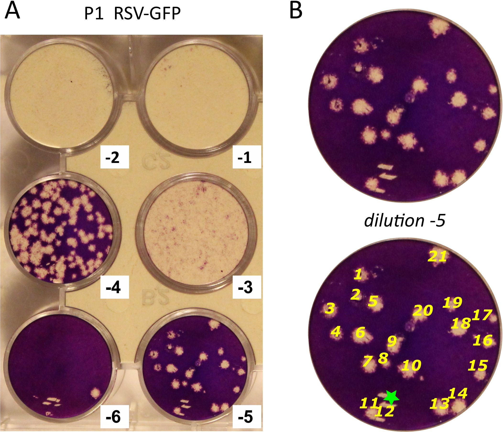
Figure 1: Determination of RSV titer using the plaque assay. (A) Results of the plaque titration assay in a 12-well plate. The six wells infected with serial dilutions of one viral stock are shown. Dilutions are indicated in a base-10 logarithm. Cells infected with the three first dilutions are all detached. The plaque numbers observed with the 10-4, 10-5, and 10-6 dilutions are consistent. (B) Illustration of the plaque enumeration (yellow numbers). The green star indicates scratches on the cell layer.
Materials
| Name | Company | Catalog Number | Comments |
|---|---|---|---|
| Crystal violet solution | Sigma | HT90132 | |
| HEp-2 | ATCC | ATCC CCL-23 | |
| MEM (10x), no glutamine | ThermoFisher Scientific | 11430030 | |
| MEM, GlutaMAX supplement | ThermoFisher Scientific | 41090-028 | |
| Sodium bicarbonate 7.5% solution | ThermoFisher Scientific | 25080094 | |
| Penicillin-Streptomycin (10,000 U/mL) | ThermoFisher Scientific | 15140122 |
