This video demonstrates adenovirus-mediated delivery of a fluorescent transgene combined with siRNA–calcium phosphate transfection in cervical cancer cells. The adenovirus expresses a fluorescent replacement protein, while the siRNA induces targeted mRNA degradation through RISC binding. This sequential co-transfection enables simultaneous knockdown of the endogenous protein and functional rescue by the transgene, allowing visualization and confirmation of protein replacement through fluorescence imaging.
Method Article
Combined Adenoviral Transduction and siRNA-Mediated Knockdown in Cancer Cells
66 views
⸱
September 30th, 2026
In This Article
Abstract
Protocol
- Adenovirus Transduction and Endogenous Protein Knockdown by Small Interfering RNA (siRNA) Transfection in HeLa-RFP-H2B Cells Using Calcium Phosphate (CaPi) Precipitates
NOTE: A control adenovirus carrying an unrelated gene (i.e., LacZ) or no gene is used to reach a minimal multiplicity of infection (MOI) in all Adenofections (10-20 plaque-forming units (PFU)/cell) using the lowest amount of recombinant adenovirus carrying the gene of interest.
NOTE: This procedure has been shown to help normalize expression per cell in a large cell population.
- One day after cell plating, count the number of cells from the additional plated dish. Aspirate the medium and rinse once with 1 mL 0.05% Trypsin/ethylenediaminetetraacetic acid (EDTA). Add again 1 mL of 0.05% Trypsin/EDTA and incubate at 37 °C, 5% CO₂ until all cells have detached. Separate the cells well using a 1 mL pipette and count the number of cells per mL using a haemocytometer.
- Determine the quantity of virus needed to transduce cells at a MOI of 2 PFU of the protein of interest (POI) per cell and 18 PFU per cell of an empty vector (e.g., LacZ) to have a total of 20 PFU per cell. At the same time, transduce 4 PFU of each baculovirus encoding green fluorescent protein (GFP)-tagged actin and red fluorescent protein (RFP)-tagged α-tubulin. Transduce the cells using the following adenofection protocol.
- Prepare in a sterile hood a 1.5 mL plastic tube for each condition and add 400 μL of warm alpha minimum essential medium (αMEM).
- Thaw an aliquot of the viruses slowly on ice and, if necessary, dilute the virus stock in order to pipet a volume greater than 1 μL to minimize pipetting errors.
- Add the calculated virus quantity per condition to each 1.5 mL plastic tube containing 400 μL αMEM and mix gently by pipetting. Place the virus stock immediately back at -80 °C to keep its activity.
- Aspirate the medium from the cells and gently pipette the virus mix dropwise. Incubate the cells at 37 °C, 5% CO₂ and agitate the plates carefully under the sterile hood every 15 min for 1 hr to ensure that the cells are well covered with the virus. Using a small amount of medium facilitates the contact of the virus with the cells.
- After incubation, gently pipette 1.6 mL αMEM to each plate to obtain a total volume of 2 mL, start cell synchronization by adding 2 mM thymidine and incubate the cells for a further 2 hr at 37 °C, 5% CO₂.
- Meanwhile, prepare the siRNA transfection mix following the CaPi transfection method (see Figure 1). If no siRNA is necessary, replace the amount of siRNA by sterile water to perform an empty transfection.
- Calculate 200 μL mix for each condition, resulting in 400 μL per duplicate. The following example is given for a final siRNA concentration of 50 nM and has to be adapted for each protein of interest. In a 1.5 mL plastic tube, pipette 50 μL 1 M CaCl2 and 11 μL 20 μM siRNA into 139 μL sterile H2O and mix by vortexing. After a quick spin, carefully add 200 μL 2× HEPES-buffered saline (HBS2x) (280 mM NaCl, 50 mM 4-(2-hydroxyethyl)-1-piperazineethanesulfonic acid (HEPES), 1.5 mM Na₂HPO₄, pH 7.01-7.05) dropwise.
NOTE: The smaller the drops, the smaller the precipitates, resulting in better transfection efficiency. Gently mix three times by air injection using a 200 μL pipette. - Incubate the mix for 30 min at room temperature (RT). Slowly add 200 μL of the transfection mix to each plate and agitate cross-wise. Transfer the plates to 37 °C, 5% CO₂ for 16 hr.
- The next day, rinse cells twice with 2 mL warm HEPES (6.7 mM KCl, 150 mM NaCl, 10 mM HEPES, pH 7.3) and add 2 mL αMEM. Do not proceed with more than four cell plates at a time since variations in temperature affect the length of the cell cycle.
- Visualize the infection efficiency under an inverted fluorescent microscope at a magnification of 20-40x (air) and acquire three representative images per condition in both fluorescent and transmission channels for documentation. Seven hours later, add 2 mM thymidine and incubate for a further 16 hr at 37 °C, 5% CO₂.
- The following day, 48 hr after siRNA transfection and virus infection, rinse the cells twice with 2 mL warm phosphate-buffered saline (PBS) and release for 7 hr in 2 mL αMEM without Phenol Red for live cell imaging or with Phenol Red for protein extraction.
- Harvest cells for each condition 48 hr post-transfection for preparation of protein extracts and Western blot analysis to determine the efficiency of knockdown and the expression of the endogenous protein.
NOTE: Use antibodies against the protein of interest as well as appropriate antibodies serving as loading controls. The efficiency of knockdown should be determined by loading decreasing amounts of control cell lysates (transfected with control siRNA) to provide a titration curve (i.e., 1, ½, ¼, ⅛).
Results
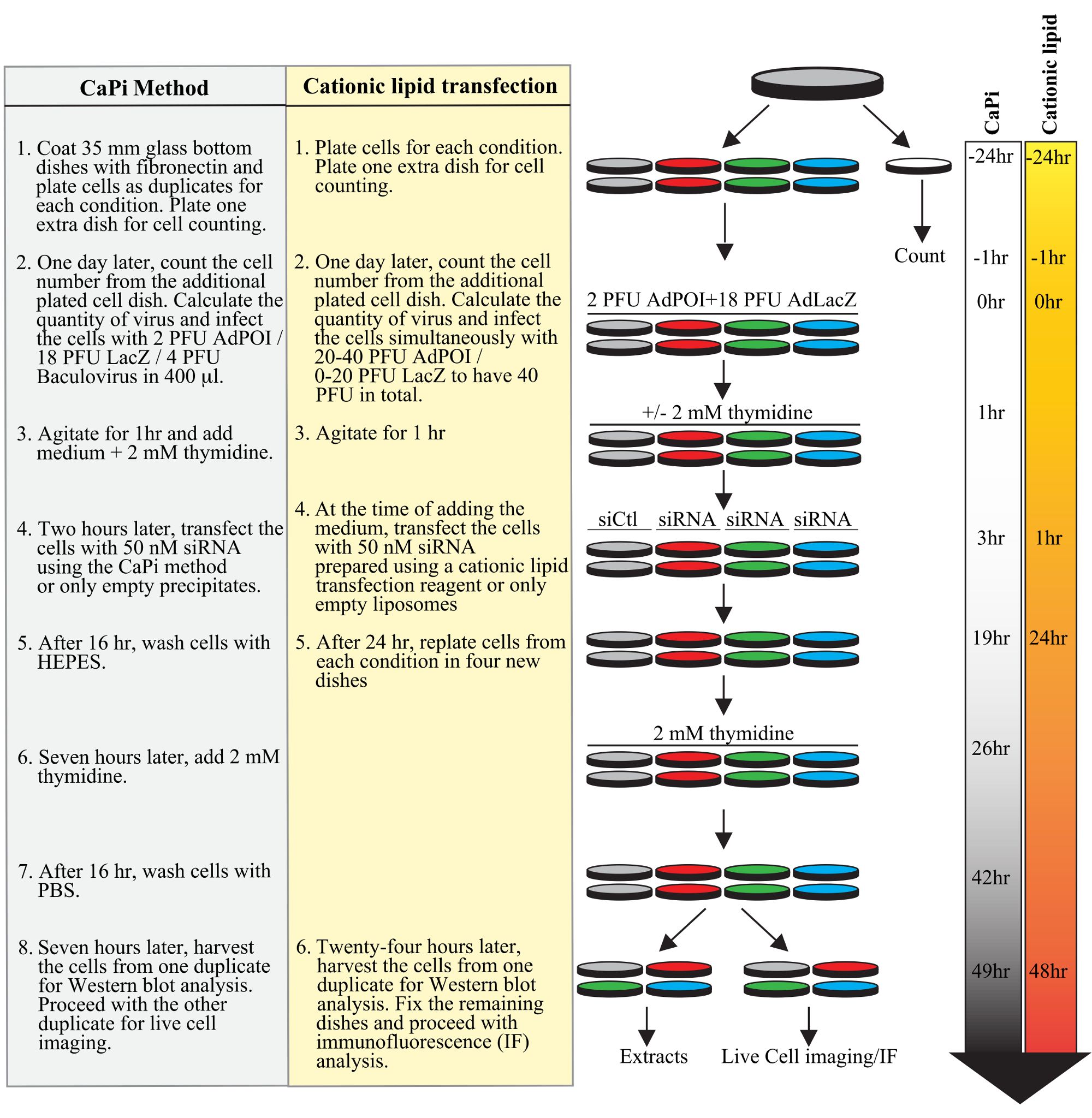
Figure 1. Planning of a typical adenofection protocol. Successive steps of a typical experiment are shown for the protocol using CaPi precipitates (underlined in grey) or the protocol using a cationic lipid transfection reagent (highlighted in yellow), including cell plating, transduction of adeno- and baculoviruses, transfection of siRNA duplexes, and cell synchronization with a double thymidine block. Time lines in hours for both protocols are shown on the right side of the figure.
Materials
| Name | Company | Catalog Number | Comments |
|---|---|---|---|
| EDTA | Sigma | E5134 | |
| Fetal Bovine Serum (FBS) | Thermo Fisher | 12483-020 | |
| Fibronectin | Sigma | F1141 | |
| Glass bottom dishes, 35 mm | MatTek Corperation | P35G-1.5-20-C Case | |
| HeLa-RFP-H2B | Kind gift of Dr Sabine Elowe, Québec, Canada | ||
| HEPES | Fisher Scientific | BP310-1 | |
| KCl | Fisher Scientific | BP366-500 | |
| L-Glutamine | Thermo Fisher | 25030081 | |
| NaCl | Fisher Scientific | BP358-10 | |
| Minimal Essential Medium (MEM) Alpha | Wisent | 310-101-CL | |
| Minimal Essential Medium (MEM) Alpha without Desoxyribonuleosides/Ribonucleosides | Thermo Fisher | 12000-022 | |
| Minimal Essential Medium (MEM) Alpha without Phenol Red | Thermo Fisher | 41061-029 | |
| Na₂HPO₄ | Biobasic | S0404 | |
| Adenovirus custom design | Welgen | Custom design | |
| Calcium Chloride | Fisher Scientific | C79-500 | |
| CellLight® Actin-GFP, BacMam 2.0 | Thermo Fisher | C10582 | |
| CellLight® Tubulin-RFP, BacMam 2.0 | Thermo Fisher | C10614 | |
| siRNA duplexes | Dharmacon | Custom design | |
| Thymidine | Sigma | T9250 | |
| Trypsine 2.5% | Thermo Fisher | 15090-046 |
