The video demonstrates the in vitro assembly of virus-specific nucleocapsids from a purified RNA-free nucleoprotein–phosphoprotein complex, their purification by size exclusion chromatography, and validation of full-length viral RNA within the assembled particles.
Method Article
In Vitro Reconstitution and Purification of Viral Nucleocapsid-Like Particles
67 views
⸱
September 30th, 2026
In This Article
Abstract
Protocol
1. Molecular cloning
NOTE: Ligase Independent Cloning (LIC) was used to make a respiratory syncytial virus (RSV) bi-cistronic coexpression construct plasmid. LIC is a method developed in the early 1990s, which uses the 3’-5’ Exo activity of the T4 DNA polymerase to create overhangs with complementarity between the vector and the DNA insert. The constructs were made using the 2BT-10 vector DNA, which consists of a 10x His tag at the N-terminal of the open reading frame (ORF) (Figure 1).
- Perform linearization of LIC vectors using SSPI digestion.
- Combine 10 μL of SSPI 10X buffer, 4 μL of SSPI enzyme at a concentration of 5 U/μL, the equivalent volume of 5 μg of vector miniprep DNA, and sterile ddH2O to 100 μL.
- Incubate the digest for 3 hours at 37 °C.
- Run the digest on a 1.0% agarose gel for the extraction of vector DNA.
- Use a gel extraction kit to perform extraction and purification. Suspend the final volume of vector DNA in 30 μL of ddH2O and store it at -20 °C.
Prepare the DNA inserts for N1-391 and P1-126 using the N1-391 Forward Primer, N1-391 Reverse Primer, P1-126 Forward Primer, and P1-126 Reverse Primer (Table 1).
NOTE: There is sufficient overlap with the linearized vector to ensure a melting temperature of between 55 °C and 60 °C. For the reverse primer, there is sufficient overlap with the reverse complementary strand of the linearized vector for the same reason.
Table 1. Primer sequences.
Primers Sequence N1-391 Forward 5’-TACTTCCAATCCAATGCAATGGCCCTGAGCAAAGTGAAG-3’ N1-391 Reverse 5’-TTATCCACTTCCAATGTTATTACAGTTCCACGTCGTTGTCCTTGG-3' P1-126 Forward 5’-TACTTCCAATCCAATGCAATGGAAAAGTTCGCCCCCGAG-3' P1-126 Reverse 5’-TTATCCACTTCCAATGTTATTACTGGTCGTTGATTTCCTCGTAGC-3’ - Perform polymerase cChain reaction (PCR) amplification of the DNA insert using the conditions in Table 2 and Table 3.
- Extract the amplified DNA insert. Run the PCR products from the previous step on a 1.0% agarose gel, then extract and purify the bands by gel extraction. Suspend the final volume of extracted DNA in 15 μL of ddH2O.
Table 2. PCR amplification of DNA insert reagents.
PCR amplification of DNA insert 15 μL 10x Pfu polymerase reaction buffer 3 μL Forward primer (100 μM concentration) 3 μL Reverse primer (100 μM concentration) 15 μL Deoxynucleotide Triphosphate (dNTP) mix at 2.5 mM concentration 6 μL Plasmid DNA contains the gene of N or P (100ng/ μL) 7 μL Dimethyl Sulfoxide (DMSO) 3 μL Pfu polymerase at 2.5U/μL Volume to fill to 150 μL Sterile ddH2O Table 3. PCR amplification of DNA insert thermocycling program.
PCR amplification of DNA insert Step Time Temperature Cycles Denaturation 4 min. 95 ºC 1 Denaturation 45 sec. 95 ºC 30 Annealing 30 sec. 62 ºC Extension* 90 sec. 72 ºC Extension 10 min. 72 ºC 1 Hold ∞ 4 ºC 1 The 150 μL mixture can be run in three separate PCR reactions (3 x 50μL). *For the Pfu DNA polymerase, 1 kb/min is the recommended speed for the extension phase. Here, both the lengths of the N1-391 gene or the P1-126 gene are shorter than 1.5 Kb. Thus, 90 seconds was used for the extension step. T4 DNA polymerase treatment of vector and insert DNA (Table 4).
Table 4. T4 DNA polymerase treatment.
T4 DNA polymerase treatment 10 x Buffer 2 μL Vector/Insert DNA (0.1 pmol vector or 0.2 pmol insert) 5 μL dNTP* at 25 mM 2 μL DTT at 100 mM 1 μL T4 DNA polymerase (LIC qualified) 0.4 μL (1.25 U) Sterile ddH2O 9.6 μL *dGTP was used for the vector, and dCTP was used for the insert DNA. NOTE: Treatment must be performed separately for the vector DNA and the insert DNA.
- Incubate the mixture for 40 minutes at room temperature. Then, heat-inactivate the polymerase at 75 °C for 20 minutes. Store the reaction mixture at -20 °C.
- Anneal the LIC vector and the insert vector.
- Set up a negative control with 2 μL of LIC vector DNA and 2 μL of sterile ddH2O.
- Combine 2 μL of insert DNA and 2 μL of LIC vector DNA from the previous T4 DNA polymerase reactions in a 0.2 mL tube.
- Perform the annealing reaction at room temperature for 10 minutes.
- Quench the reaction with 1.3 μL of Ethylenediaminetetraacetic Acid (EDTA) at a concentration of 25 mM.
- Transform the reaction into 100 μL of Escherichia coli Top10 competent cells and plate them on an ampicillin selection plate.
- Identify the positive constructs.
- Prepare the plasmid miniprep solution. This can be done through colony picking and inoculation in Luria Broth (LB) media. Usually, 3 colonies are sufficient.
- Incubate the mixture overnight at 37 °C.
- Centrifuge the mixture at 4,560 x g for 10 minutes and discard the supernatant.
- Resuspend the pellets in 250 μL of P1 buffer and prepare plasmid minipreps using a spin miniprep kit.
- Conduct a digestion analysis of the miniprep product using AseI or other restriction enzymes.
- Load samples on 0.8% agarose gel and run the digested plasmid. Analyze the gel under a UV lamp.
- Use a sequencing service to validate the sequence of the positive product.
Obtain the coexpression DNA insert.
- Perform PCR to obtain N1-391 and P1-126 using the previously constructed 2BT-10 N1-391 and 2BT-10 P1-126 as templates.
- Perform the 1st PCR to obtain N1-391 from the 2BT-10 N1-391 construct using the PCR conditions in Table 2 and Table 3 with the N1-391 Forward primer and Reverse Primer 5 ́-GTGAAGATCCTGGCTGATGCAATGCGGCGGCGCGCCGCGATCGCGGATCC-3 ́.
- Perform the 2nd PCR to obtain P1-126 from the 2BT-10 P1-126 construct using the Forward primer: 5’-CCGCCGCATTGCATCAGCCAGGATCTTCACTGCAGGACTCGAGTTCTAGA-3 ́and the P1-126 Reverse primer (use the PCR conditions in Table 2 and Table 3).
- Finally, perform overlap PCR on the mixed products of the previous 2 PCR reactions to merge N1-391 and P1-126. Use the N1-391 Forward primer and P1-126 Reverse primer. Use the PCR conditions in Table 5 and Table 6.
Table 5. Overlap PCR reagents.
Overlap PCR 15 μL 10 X Pfu polymerase reaction buffer 3 μL Forward primer (100 μM) 3 μL Reverse primer (100 μM) 15 μL dNTP Mix (2.5 mM) 3 μL DNA from 1st round PCR which contain the gene N1-391 (100 ng/μL ) 3 μL DNA from 1st round PCR which contain the gene P1-126 (100 ng/μL ) 7 μL DMSO 3 μL Pfu polymerase (2.5 U/μL) Volume to fill to 150 μL Sterile ddH2O Table 6. Overlap PCR thermocycling program.
Overlap PCR Step Time Temperature Cycles Denaturation 4 min. 95 °C 1 Denaturation 45 sec. 95 °C 30 Annealing 30 sec. 62 °C Extension* 2 min. 72 °C Extension 10 min. 72 °C 1 Hold ∞ 4 °C 1 The 150 μL mixture can be run in three separate PCR reactions (3 x 50 μL). *For the Pfu DNA polymerase, 1 kb/min is the recommended speed for the extension phase. Here, the total length of the gene N1-391 and P1-126 is shorter than 2.0 Kb. Thus, 2 minutes were used for the extension step. - Join the vector and the DNA insert.
- Treat the overlap PCR product with T4 DNA polymerase following the protocol in step 1.3.
- Anneal the LIC vector and PCR product following the protocol in step 1.4.
- Identify the positive constructs following the protocol in step 1.5.
2. Protein expression and purification
NOTE: Use E. coli for the bi-cistronic construct of the coexpression of both N and P. Culture the cells at 37 °C, but carry out the expression at a reduced temperature (16 °C) overnight. Purify the protein complexes through a combination of cobalt column, ion exchange, and size exclusion chromatography (Figure 2).
- Use the E. coli BL21(DE3) strain for protein production. Grow 4 L cell cultures at 37 °C in LB (Luria Broth) medium until OD600 reaches 0.6.
- Lower the temperature to 16 °C. An hour later, induce the expression with 0.5 mM Isopropyl β-D-1-thiogalactopyranoside (IPTG) overnight.
- Centrifuge the cells at 4,104 x g for 25 min and then discard the supernatant.
- Resuspend the cell pellets in 200 mL of lysis buffer A (50 mM sodium phosphate, pH 7.4, 500 mM NaCl, 5 mM imidazole, 10% glycerol, and 0.2% NP40). Use 50 mL of lysis buffer to resuspend the cell pellets from 1 L of cell culture.
Lyse the cells by sonication for 15 min, 3 seconds on, and 3 seconds off. Then centrifuge cells at 37,888 x g for 40 min.
NOTE: The protocol can be paused by freezing the cells before sonication in a -80 °C freezer.
- Load the supernatant into a cobalt gravity column (diameter x length: 2.5 cm x 10 cm) with ~10mL of beads pre-equilibrated with 5-10 column volumes (CV) of lysis buffer.
- Wash the column with 5 CV of buffer B (50 mM Tris-HCl pH 7.4, 1 M NaCl, 10% glycerol, and 5 mM imidazole) and 5 CV of buffer C (50 mM Tris-HCl pH 7.4, 500 mM NaCl, 10% glycerol, and 5 mM imidazole).
- Elute the protein from the beads using 2 CV of buffer D (50 mM Tris-HCl pH 7.4, 500 mM NaCl, and 250 mM imidazole).
- Dilute the eluted protein 5x with QA buffer (50 mM Tris-HCl pH 8.0, 5% Glycerol) for the Q column.
- Wash the 5 mL of Q column with QA buffer to equilibrate the column, then load the diluted sample into the Q column using the peristaltic pump (e.g., Rabbit).
- Load the Q column into the HPLC machine along with QA buffer and QB buffer (50 mM Tris-HCl pH7.4, 1.5 M NaCl, 5% glycerol). Set up the flow rate as 1 mL/min.
- Run the “pump wash” program to wash the machine with QB buffer followed by QA buffer (1-2 CVs/each). Set the system flow to 3 mL/min.
- Set the UV1 to 280 nm and UV2 to 260 nm. Use a 96 deep-well plate to collect the fractions.
- Elute proteins using a stepwise gradient of elution agent (QB Buffer) applying 3-4 CV of each concentration, increasing the percentage by 5% each time starting at 0% QB. N0P protein complex will come out at 15% QB Buffer.
- Once all of the protein is eluted, wash the column with 100% QB Buffer (2 CV).
- Isolate the protein by gel filtration Superdex 200 Increase 10/300 GL column (diameter x length: 1.0 cm x 30 cm) and equilibrate with buffer E (20 mM 4-(2-hydroxyethyl)-1-piperazineethanesulfonic acid or, HEPES pH 7.4 and 200 mM NaCl).
- Analyze protein-containing fractions by sodium dodecyl sulfate polyacrylamide gel electrophoresis (SDS-PAGE).
3. In vitro assembly of the virus-specific NC
NOTE: The in vitro assembly of the RSV-specific NC (N:RNA) was performed by incubating the prepared N0P complex with RNA oligos. Then, size exclusion chromatography (SEC) was used to separate the assembly complex from the N0P and excess RNA (Figure 2).
- Mix and incubate the purified N0P complex with RNA oligo with the molecular ratio of 1:1.5 at room temperature for 1 hour, usually 1 mL of the protein N0P with a concentration of 1 mg/mL is enough for the next step. Set up the control sample, which only contains the same amount of N0P protein.
- Pre-equilibrate the gel filtration Superdex 200 Increase 10/300 GL column with the buffer E (20 mM HEPES, pH 7.4, 200 mM NaCl).
- Centrifuge the sample with 21,130 x g for 15 min, remove any precipitation and load the supernatant to the SEC column.
- Compare the SEC chromatography images of N:RNA assembly sample and N0P control sample, combine the A260/A280 ratio to identify which peaks are the assembled N-RNA, N0P, and free RNA.
- Collect the peak fractions, run the SDS-PAGE gel, or make grids.
- For the assembly N-RNA complex, collect all the fractions of N-RNA peak, do the RNA extraction and run the Urea-PAGE gel to double-check the length of specific RNA, which is the same as mixed and incubated at the first step.
Results
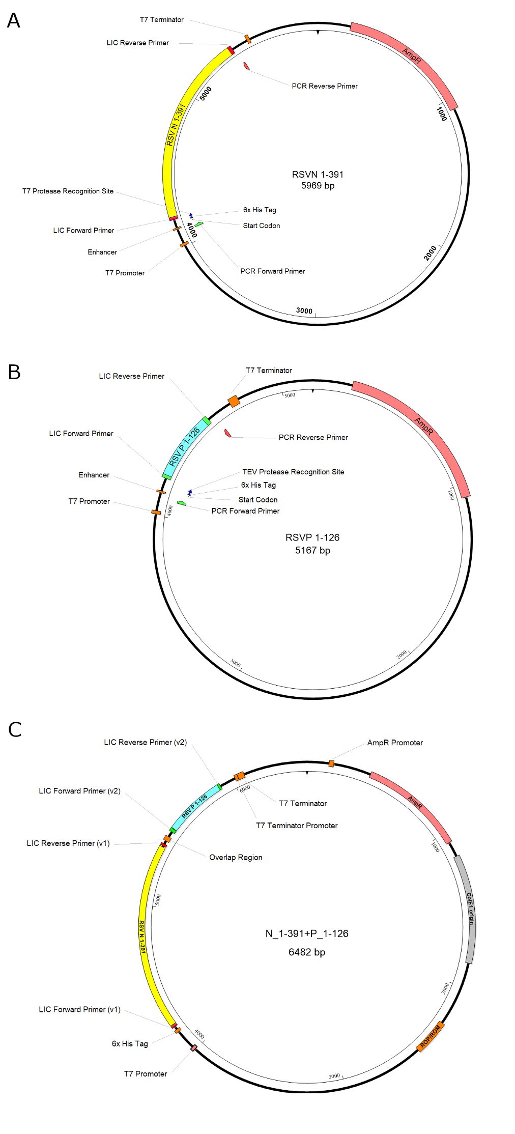
Figure 1. The illustration of the plasmid constructions. A. The construct of the RSV N1-391; B. The construct of the RSV P1-126; C. The bi-cistronic construct for the coexpression of N1-391 and P1-126. The first gene RSV N1-391, the second gene RSV P1-126, the antibiotic-resistant gene (AmpR), and the promoters are highlighted in yellow, cyan, pink, and orange boxes, respectively. In summary, the gene inserts of the RSV N1-391 and the RSV P1-126 are constructed separately and assembled.
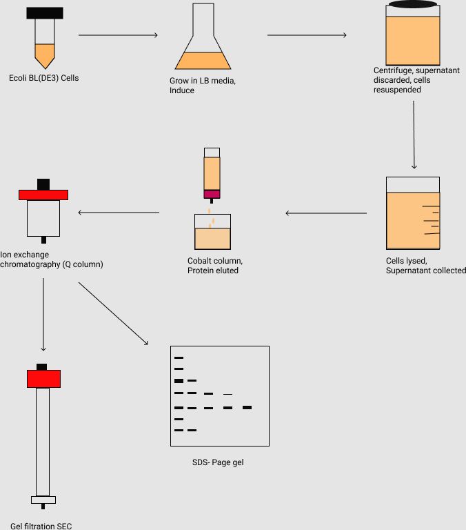
Figure 2. The flowchart of the purification of the protein complex N1-391P1-126. It outlines the inoculation and large scale grow-ups of the E. coli cell culture and harvesting the cell by centrifugation. Followed by cell lysis, the protein samples are purified by the affinity chromatography (i.e., Co2+ column), ion-exchange chromatography (i.e., Q column), and gel filtration size exclusion (SEC) chromatography. The protein samples are further analyzed by the SDS-PAGE gel.
Materials
| Name | Company | Catalog Number | Comments |
|---|---|---|---|
| Agarose | SIgma | A9539-500G | making construct using LIC method |
| Ampicillin sodium | GOLD BIOTECHNOLOGY | 5118.111317A | antibiotic for cell culture |
| AseI | NEB | R0526S | making construct using LIC method |
| Cobalt (High Density) Agarose Beads | Gold Bio | H-310-500 | For purification of His-tag protein |
| Corning LSE Digital Dry Bath Heater | CORNING | 6885-DB | Heat the sample |
| dCTP | Invitrogen | 10217016 | making construct using LIC method |
| dGTP | Invitrogen | 10218014 | making construct using LIC method |
| Glycerol | Sigma | G5516-4L | making solution |
| HEPES | Sigma | H3375-100G | making solution |
| HiTrap Q HP | Sigma | GE29-0513-25 | Protein purification |
| Imidazole | Sigma | I5513-100G | making solution |
| IPTG (Isopropyl-beta-D-thiogalactopyranoside) | GOLD BIOTECHNOLOGY | 1116.071717A | induce the expression of protein |
| Microcentrifuge Tubes | VWR | 47730-598 | for PCR |
| Misonix Sonicator XL2020 Ultrasonic Liquid Processor | SpectraLab | MSX-XL-2020 | sonicator for lysing cell |
| New Brunswick Innova 44/44R | eppendorf | M1282-0000 | Shaker for culturing the cell |
| Nonidet P 40 Substitute | Sigma | 74385-1L | making solution |
| OneTaq DNA Polymerase | NEB | M0480L | PCR |
| QIAquick Gel Extraction Kit | QIAGEN | 28706 | Purify DNA |
| SSPI-HF | NEB | R3132S | making construct using LIC method |
| Superose 6 Increase 10/300 GL | Sigma | GE29-0915-96 | Protein purification |
| T4 DNA polymerase | Sigma | 70099-3 | making construct using LIC method |
| Thermo Scientific Sorvall RC 6 Plus Centrifuge | Fisher Scientific | 36-101-0816 | Centrifuge, highest speed 20,000 rpm |
| Trizma hydrochloride | Sigma | T3253-250G | making solution |
