The video demonstrates the detection of Zika viral dsRNA intermediates in infected epithelial cells using immunocytochemistry. Infected cells are fixed and then blocked with a blocking solution. The primary and secondary antibodies are added. The nuclei are stained with a fluorescent dye. The labeled viral dsRNA is then seen under a fluorescence microscope.
Method Article
Detection of Zika Viral dsRNA Replication Intermediates Using Immunocytochemistry
80 views
⸱
September 30th, 2026
In This Article
Abstract
Protocol
1. Zika viral genome replication assay
- Seed naïve Vero cells at 1 x 10⁵ cells per well in 2 ml volumes using a 12-well plate. The following day, prepare viral inocula (Multiplicity of Infection (MOI) of 0.01 and 0.1; 400 µl/well) of low and high titer using serum-free media in triplicate. For mock infection, use serum-free media (400 µl/well) only.
- Incubate the inoculated plate at 37 °C with 5% CO2 for 4-6 hr. Spread the inoculum by gently tilting the plate sideways every one hr.
- At the end of incubation, replace the inoculum with serum-supplemented media (2 ml per well).
- At the 48 and 96 hr time points, collect the samples for immunocytochemistry (ICC).
- Perform ICC using the methanol-fixed cells.
- Wash the fixed cells three times with 1x phosphate-buffered Saline (PBS) and block with ICC blocking buffer (3% goat serum, 3% Bovine serum albumin (BSA), 0.1% Triton-x 100 in PBS).
- Use mouse monoclonal anti-dsRNA antibody J2 (1 µg/ml) at a dilution of 1:100 in blocking buffer and incubate overnight at 4 °C.
- Wash the cells with 1x PBS and add a secondary antibody goat anti-mouse IgG-594 (1 µg/ml) at a 1:1,000 dilution in the blocking buffer and incubate for one hour at room temperature.
- Wash cells with 1x PBS and stain for nuclei using Hoechst dye and observe the cells using a fluorescent microscope at 100X magnification (Figure 1).
Results
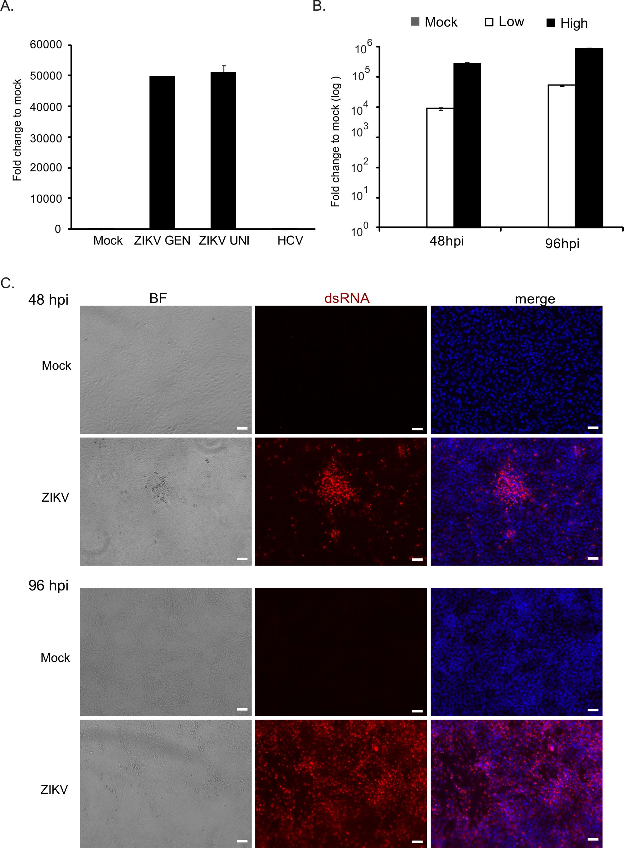
Figure 1: Assays for evaluating Zika virus (ZIKV) replication. (A) The graph shows Zika virus genomic content measured by Reverse Transcription Quantitative Polymerase Chain Reaction (RT-qPCR). Both pan-genotype (ZIKV GEN) and PRVABC59-strain (ZIKV UNI) primers showed equal sensitivity and specificity. As expected, hepatitis C viral (HCV) primers did not amplify any product. (B) The graph presents the Zika viral growth kinetics at the indicated time points in low and high-titer infected cells compared to that of the uninfected mock control. Mean values and standard deviations are given. (C) Immunocytochemistry assay for investigating viral replication. Zika virus-infected cells were specifically stained with dsRNA antibodies. At 48 hpi, infected cells are in a few clusters, whereas at 96 hpi, most of the cells are infected. Hoechst stain was used for the visualization of nuclei (Scale bar = 50 µm). BF: Bright field image.
Materials
| Name | Company | Catalog Number | Comments |
|---|---|---|---|
| Dulbecco’s modified Eagle’s medium (DMEM) | Sigma Life Science | D5796 | |
| HEPES | Life Technologies | 15630080 | |
| Glutamax | Life Technologies | 35050061 | |
| Mouse monoclonal anti-dsRNA antibody J2 | English & Scientific Consulting Kft. | 10010200 | |
| Goat anti-rabbit IgG Alexa Fluor 594 | Life Technologies | A11020 | |
| Vero Cell Line | ATCC | CCL-81 | |
| Zika viral strain PRVABC59 | Centers for Disease Control and Prevention (CDC) | ||
| Nikon Eclipse Ti Immunofluorescence Microscope with Nikon Intenselight C-HGFI | Nikon | Visit Nikon for Request |
