All procedures involving human participants have been performed in compliance with the institutional, national, and international guidelines for human welfare and have been reviewed by the local institutional review board.
1. Isolation and Culture of Primary Untransformed Stromal Cells
- Human tissue collection and preparation.
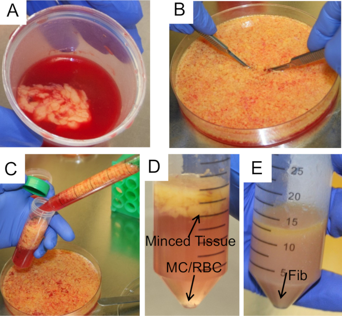
- Obtain specimens of human omentum, 2 cm3, removed during an abdominal surgery, and immediately immerse tissue in RT phosphate-buffered saline (PBS) (Figure 1A). A piece of omentum 3 cm x 2 cm typically yields 1 million primary human mesothelial cells (HPMC) and 200,000 primary human fibroblasts or normal omental fibroblasts (NOF).
- Spin down the PBS the tissue was immersed in at 0.5 x g for 3 min as soon as possible after collection (< 2 hr in PBS) and transfer the tissue to fresh 20 ml PBS in a 50 ml conical tube. Cut up tissue into 5mm3 pieces by mincing with scalpels in a 15 cm diameter, sterile culture dish (Figure 1B).
- Primary human mesothelial cell isolation
- To isolate HPMC, transfer the minced tissue into 50 ml conical tubes and wait 1 min for the solid pieces to float to the top (Figure 1C & D). HPMC and red blood cells (RBC) will be present in the liquid on the bottom. Use a pipette to remove the liquid from the bottom of the tube and into a new conical tube. Spin the liquid down at 0.5 x g for 3 min and aspirate the supernatant from the HPMC/RBC pellet.
- Repeat the process three times: add 20 ml PBS to the minced tissue, allow tissue to rise, remove liquid from the bottom with a pipette and into the HPMC/RBC tube, spin down, and remove the supernatant. After all spins are completed and the supernatant is aspirated, the pellet will contain HPMC and RBC.
- Plate all cells from the pellet into a 75 cm2 flask in 15 ml of full growth media (DMEM with 10% fetal bovine serum [FBS],1% MEM vitamins [93 mg/L], 1% MEM nonessential amino acids [81.4 mg/L], 1% penicillin streptomycin [Pen-Strep, 100 U/ml penicillin and 100 µg/ml streptomycin]).
- Perform a secondary PBS wash to isolate additional HPMCs.
- For the secondary PBS wash, shake the remaining solid tissue at 200 rpm, 37 °C for 10 min in 20 ml of PBS, then wait 1 min for the tissue to float to the top. Remove the liquid from the bottom of the tube into a new tube, centrifuge at 0.5 x g for 3 min, and aspirate the supernatant.
- Plate all the HPMC/RBC from the secondary PBS wash in a separate 75 cm2 flask in 15 ml of full growth media.
- To isolate any remaining HPMC, shake the minced tissue at 200 rpm, 37 °C degrees for 10 min in 20 ml of a 1:1 0.25% trypsin 25 mM EDTA:PBS solution by volume. Allow the tissue to rise, collect the liquid at the bottom of the tube into a new 50 ml tube, and spin down at 0.5 x g for 3 min.
- Aspirate the supernatant and plate all the HPMC/RBC in a separate 75 cm2 flask in 15 ml of full growth media.
- Culture the plated cells at 37°C and 5% CO2 in a humidified environment. Feed plated cells by adding 15 ml of full growth media on days 3 and 5 without removing the spent media. HPMC can be cultured for 5-7 days before splitting.
- Use low-passage HPMC (up to passage 2) in all experiments to minimize dedifferentiation and modification of original phenotype. Use a wide-field microscope preferably with a 20x objective with plastic differential interference contrast capabilities or 4-20x objective with phase contrast capabilities (phase contrast filters on microscope) for taking all images of cells, and a 10x objective ()in bright field to analyze immunohistochemical 3,3'-Diaminobenzidine (DAB) staining.
- Confirm that the HPMC are cuboidal and express cytokeratin 8 and vimentin by performing immunohistochemistry (Figure 2A, C, E).