Source: Shechtman, L. A. et al. Generation and Culture of Lingual Organoids Derived from Adult Mouse Taste Stem Cells. J. Vis. Exp. (2021)
In this video, we describe the isolation of circumvallate papillae (CVP) epithelium from a murine tongue.
A subscription to JoVE is required to view this content. Sign in or start your free trial.
Method Article
4.1K views
⸱
July 8th, 2025
Source: Shechtman, L. A. et al. Generation and Culture of Lingual Organoids Derived from Adult Mouse Taste Stem Cells. J. Vis. Exp. (2021)
In this video, we describe the isolation of circumvallate papillae (CVP) epithelium from a murine tongue.
All procedures involving animal models have been reviewed by the local institutional animal care committee and the JoVE veterinary review board.
1. Isolation of CVP Epithelium
Access restricted. Please log in or start a trial to view this content.
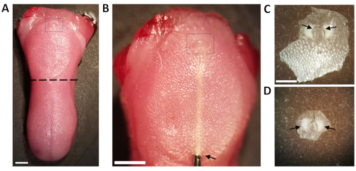
Figure 1: Dissected tongue and peeled CVP epithelium. (A) The tongue is dissected out, and the anterior tongue is removed by cutting ju.......
Access restricted. Please log in or start a trial to view this content.
| Name | Company | Catalog Number | Comments |
|---|---|---|---|
| 1 mL TB Syringe | BD Syringe | 309659 | |
| Dulbecco’s Phosphate Buffered Saline with Ca²? & Mg²? | Gibco | 10010-023 | |
| Dulbecco’s Phosphate Buffered Saline with Ca²? & Mg²? | Sigma Life Sciences | D8662-500ML | |
| Dumont #5 Forceps | Fine Science Tools | 11252-30 | |
| Fetal Bovine Serum (FBS) | Gibco | 26140-079 | |
| 1 mL TB Syringe | BD Syringe | 309659 | |
| 2.0 mL Microcentrifuge Tubes | USA Scientific | 1420-2700 | |
| Surgical Scissors - Sharp | Fine Science Tools | 14002-14 | |
| Extra Fine Bonn Scissors | Fine Science Tools | 14084-08 | |
| Big-Digit Timer/Stopwatch | Fisher Scientific | S407992 | |
| Stereomicroscope | Zeiss | Stemi SV6 | |
| Cohan-Vannas Spring Scissors | Fine Science Tools | 15000-02 | |
| Collagenase from Clostridium histolyticum, type I | Sigma Life Science | C0130-1G | |
| Dispase II (neutral protease, grade II) | Sigma-Aldrich (Roche) | 4942078001 |
