Method Article

Isolation of Mouse Valve Interstitial Cells: An Enzymatic Method to Isolate and Culture VICs from Mouse Aortic Valve

July 8th, 2025

In This Article

Abstract

Loading...
$$\rightleftharpoonup{xx}$$ $$\longleftharp{xx}$$, $$\longrightharp{xx}$$,

Source: Bouchareb, R. et al. Isolation of Mouse Interstitial Valve Cells to Study the Calcification of the Aortic Valve In Vitro. J. Vis. Exp. (2021)

In this video, we demonstrate the isolation of aortic valve interstitial cells from the heart of an adult mouse using a two-step collagenase procedure and their subsequent culture in low volume of growth media.
Protocol

Loading...
$$\rightleftharpoonup{xx}$$ $$\longleftharp{xx}$$, $$\longrightharp{xx}$$,

All procedures involving animal models have been reviewed by the local institutional animal care committee and the JoVE veterinary review board.

1. Preparation Before Valve Cell Isolation from Adult Mice

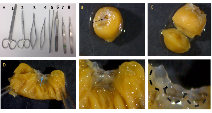
  1. Clean and sterilize all the surgical instruments shown in Figure 1A by using 70% v/v ethanol and subsequently autoclaving them for 30 min. Clean the surgical workspace with 70% ethanol.
  2. Add 500 µL of penicillin-streptomycin to 50 mL of 10 mm HEPES. Prepare an aliquot of 50 mL of 1x phosphate-buffered saline (PBS). Keep the solutions on ice.
  3. Prepare 1 mg/mL and 4.5 mg/mL collagenase solutions, and use 5 mL of each solution in 15 mL tubes to perform the entire procedure. To prepare 5 mL of 1 mg/mL collagenase, mix 5 mg of collagenase with 2.5 mL of Dulbecco's Modified Eagle Medium (DMEM, fetal bovine serum (FBS)-free) and 2.5 mL of 10 mM HEPES supplemented with antibiotics (1% penicillin-streptomycin from step 1.2). Filter the solutions through a 0.22 µm filter to remove any contamination.
    NOTE: Keep the solutions on ice to protect the enzymes.
  4. Warm the DMEM solution to 37 °C before use in all the steps described below. Prepare complete medium by supplementing DMEM with 1% penicillin-streptomycin, 1% sodium pyruvate, 5 mL of 200 mM L-glutamine, 1 mL of mycoplasma elimination reagent (see the Table of Materials), and 10% FBS.

2. Isolation of Valve Cells

  1. To obtain 106 cells for the experiment, use five 8-week-old mice (minimum of three). Place the mouse in an induction chamber along with a small piece of tissue paper soaked with 1 mL of isoflurane, but do not allow contact with the tissue. To confirm that the animal is fully anesthetized; check for toe pinch reflex, and then euthanize the mouse by cervical dislocation. Use isoflurane to alleviate any pain prior to the cervical dislocation as the procedure described below is terminal.
  2. Place the mouse on a dissecting platform, and fix the paws with cannulas to hold it in place. Clean the chest and the abdomen with ethanol; open the abdomen and the chest with scissors. With small surgical scissors, cut between the left atrium and the left ventricle to exsanguinate the mouse. Perfuse the heart with 10 mL of cold 1x PBS to remove blood from the heart.
  3. Cut the heart, and keep 3 mm from the ascending aorta as shown in Figure 1B. Dissect the aortic valve under a stereomicroscope. Cut the heart horizontally in the middle of the ventricles (Figure 1C). Cut the left ventricle toward the aorta, and carefully dissect the aortic valve (Figure 1D-F). Pool the valves together in a small 35 mm tissue culture dish.
  4. Wash the isolated valves in a 75 mm cell culture dish with 5 mL of cold HEPES (10 mM) supplemented with antibiotics (1% penicillin-streptomycin) to remove blood (Figure 2). Prepare two 15 mL tubes of collagenase 1 mg/mL and 4.5 mg/mL as described above in step 1.3.
    NOTE: After the dissection, manipulate the isolated valves in a sterile biosafety hood to minimize contamination.
  5. Incubate the valves in collagenase type I (1 mg/mL) for 30 min at 37 °C with continuous shaking (Figure 2). Centrifuge the tube for 5 min at 150 × g, wash the pellet once with 2 mL of HEPES (10 mM), and vortex for 30 s at high speed. Pour the contents of this tube into a 35 mm culture dish, and carefully transfer the fragments of tissue using thin tweezers into a new tube.
    NOTE: At this stage, the VICs are still not dissociated from the tissue, and the pellet contains pieces of tissue. To avoid contamination with endothelial cells, do not centrifuge after vortexing in step 2.5.
  6. Incubate the pellet in a 15 mL tube with 5 mL of collagenase type I (4.5 mg/mL) at 37 °C under continuous agitation for 35 min. Re-suspend the cells with a 1 mL pipette to separate the cells, and centrifuge at 150 × g for 5 min at 4 °C.
  7. Discard the supernatant, and re-suspend the pellet in 2 mL of complete DMEM. Centrifuge at 150 × g for 5 min at 4 °C. Repeat this step twice to clean the cells.
    NOTE: The pellet will still have some tissue fragments.
  8. Re-suspend the pellet in 1 mL of complete medium, and plate the cells in one well of a 6-well cell culture dish in a minimum amount of medium to facilitate their attachment to the culture dish. Leave the cells, undisturbed, in a 37 °C incubator with 5% carbon dioxide.
  9. After 3 days, check the cells under the microscope to verify good growth close to the tissue debris. Once 1,000 cells are visible under the microscope, carefully remove the tissue debris with autoclaved tweezers, and change the medium.
    NOTE: The plate should not be disturbed; if the required number of cells are not observed, place the cell culture dish back in the incubator for another 2 days.

Access restricted. Please log in or start a trial to view this content.

Results

Loading...
$$\rightleftharpoonup{xx}$$ $$\longleftharp{xx}$$, $$\longrightharp{xx}$$,

Dissection process with surgical tools and chickpea seed dissection, microscopic tissue analysis.
Figure 1: Description of valve dissection. (A) Representative image of all the surgical instruments needed for the dissection, scissors 2 is needed to open the skin of the mouse and scissor...

Access restricted. Please log in or start a trial to view this content.

Disclosures

Loading...
$$\rightleftharpoonup{xx}$$ $$\longleftharp{xx}$$, $$\longrightharp{xx}$$,
No conflicts of interest declared.

Materials

List of materials used in this article
NameCompanyCatalog NumberComments
3 mm cutting edge scissorsF.S.T15000-00
Bonn ScissorsF.S.T14184-09
Collagenase type I (125 units/mg)Thermofisher Scientific17018029
DMEMThermofisher11965092
Extra fine graefe forcepsF.S.T11150-10
FBSGibco 16000044
Fine forcepsF.S.T Dumont
HEPES 1 M solutionSTEMCELLS TECHNOLOGIES
L-Glutamine 100xThermofisher ScientificA2916801
MycozapLanzaVZA-2011Mycoplasma elimination reagent
PBS 10xSIGMA-ALDRICH
Pencillin streptomycin 100xThermofisher Scientific10378016
Sodium Pyruvate 100 mMThermofisher Scientific11360070
Standard pattern forcepsF.S.T11000-12
Surgical Scissors - Sharp-BluntF.S.T14008-14
Trypsin 0.05%Thermofisher Scientific25300054

Reprints and Permissions

Request permission to reuse the text or figures of this JoVE article

Request Permission

Tags

Aortic Valve IsolationCollagenase DigestionCell Culture ProtocolEnzymatic DissociationValve Leaflet DissectionCentrifugation ProcedureLow Volume PlatingHEPES BufferComplete DMEM

Related Articles