All procedures involving animal models have been reviewed by the local institutional animal care committee and the JoVE veterinary review board.
1. Pre-operation preparation
- Instrument sterilization: Steam-sterilize surgical instruments in an autoclave (121 °C for 15 min) prior to the surgery. Pack instruments in a metal container and maintain them until they are used in the surgery.
- Surgery platform setup: Assign a bench area of at least 60 cm x 60 cm for the operation. Clean the surface of area with 75% alcohol and cover with a disposable medical towel. Place a sterile surgical instruments pack, reagents, surgical items onto a disposable medical towel within the upper 1/3 of area. Leave the remaining 2/3 of area clean for surgical operation. Add a hotpad underneath surgical pad for thermal support.
- Animal preparation
- Place the animal (C57BL/6J mice, male, 8-week old) into the induction chamber. Turn on the vaporizer at an induction level of 4% for isoflurane and 4 L/min for oxygen. After the animal is fully anesthetized, maintain the anesthetic with the nose cone and the anesthetic delivery at a level of 1.5% for isoflurane and 0.4 L/min for oxygen during surgery. Monitor the animal for respiration.
- Apply chlortetracycline hydrochloride eye ointment to prevent corneal dryness during the surgery.
- Shave the surgical area on the dorsal surface from the lower thoracic region to the top of the sacral region using a small animal trimmer. Remove the shaved fur with tissue wipes.
- Apply depilatory cream onto the shaved area and leave it there no longer than 3 min. Remove the cream with gauze and flush with 2 mL of 0.9% sterile saline.
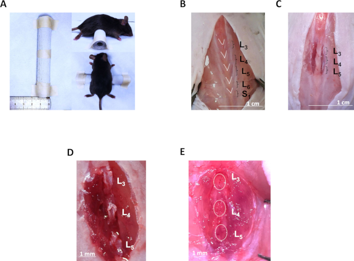
- Place a custom-made surgical cylindrical pad (Figure 1A) under the abdomen of the mouse to raise up the lumbar spine and facilitate the surgical operation.
2. Exposure of the lumbar third to lumbar fifth (L3–L5) spinous processes
- Use the index finger to touch the subcutaneous spinous processes of the lumbar vertebrae, which are more outward, and compare with thoracic vertebrae and sacral vertebrae to identify the lumbar region.
- Rinse the skin using 75% alcohol. Perform a 3–4 cm midline skin incision over the lumber region from the mid-thoracic region to the hip using a scalpel blade to expose the fascia.
- Identify the lumbar spine by the morphology of the posterior fascia inserted onto the tips of the spinous processes. In detail, the third lumbar (L3) to the first sacral (S1) fasciae are distinct from other fasciae by their “V” shapes. The last “V” tip connects to the first sacral (S1) fascia and the first “V” tip corresponds to the L3 spinous process (Figure 1B).
- Make the posterior paraspinous muscle incisions along the spinous processes from L3 to L5 on both sides laterally with a scalpel blade (Figure 1C). Control the incision depth towards the facets to reduce hemorrhage.
- Separate the muscle layers using two ophthalmic forceps to expose L3 to L5 spinous processes and supraspinous ligaments.
3. Resection of L3–L5 spinous processes along with the ligaments
- Separate individual spinous processes by cutting off interspinous ligaments using Venus shears (Figure 1D).
- Resect the L3–L5 spinous processes along with the interspinous ligaments with Venus shears (Figure 1E).
- Suture the skin incision with sterile silk braided (suture size 5.0) without reattachment of the paravertebral muscles.
- Apply Chlortetracycline Hydrochloride Eye Ointment to the surgical site.