A subscription to JoVE is required to view this content. Sign in or start your free trial.

Method Article

Hepatic Angiography: A Procedure to Visualize Liver Tumor Vessels in a Rabbit Model

3K views

July 8th, 2025

In This Article

Abstract

Source: Khabbaz, R. C., et al. Development and Angiographic Use of the Rabbit VX2 Model for Liver Cancer. J. Vis. Exp. (2019).

In this video, we demonstrate a step-by-step procedure to perform hepatic angiography using a fluoroscope that enables the real-time visualization of blood vessels associated with a liver tumor in a rabbit model.
Protocol

All procedures involving animal models have been reviewed by the local institutional animal care committee and the JoVE veterinary review board.

1. Angiographic Utilization of the VX2 Liver Tumor

  1. Anesthetize the rabbit using 45 mg/kg ketamine and 5 mg/kg xylazine for induction followed by intubation and maintenance with 1%-3% isoflurane as needed. When the rabbit is anesthetized, shave the surgical site using hair clippers. Clean the incision area using a triplicate wash with betadine scrub, 70% ethanol solution, and betadine solution, and drape the groin area in a sterile surgical fashion.
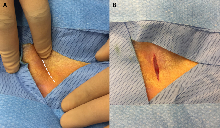
  2. Palpate the femoral groove in the groin. When the groove has been identified, make a 2-3 cm linear incision along the groove (Figure 1).
  3. Using blunt dissection, locate and isolate the femoral bundle containing the femoral vein, artery, and nerve (Figure 2).
  4. Again, use blunt dissection to separate the femoral artery from the rest of the structures in the bundle and isolate the artery atop a scalpel handle (Figure 3).
  5. With a 3-French introducer kit, utilize the Seldinger technique to gain access with a needle. Insert a guidewire and remove the needle to advance the 3-French sheath into the vessel (Figure 4).
    CAUTION: Avoid using excessive force in advancing the 3-French sheath, as this can result in transection of the femoral artery.
  6. Under fluoroscopic guidance and using a catheter, guidewire, and iohexol contrast agent, select for the celiac trunk — typically located at the T12 level — and then advance the catheter into the left hepatic artery via the common hepatic and proper hepatic.
  7. At this point, administer the agent of choice through the catheter. Once the agent is administered, remove the catheter.
  8. Using 3-0 silk suture, ligate the femoral artery proximally and distally to the insertion point of the sheath. Be sure to tighten the knot proximal to the sheath as it is withdrawn to prevent bleeding.
  9. Close the groin incision with 4-0 polyglactin 910 sutures on a cutting needle using a subcuticular stitch.
  10. Maintain standard post-operative care and monitor animal recovery. Perform euthanasia and necropsy as needed using standard techniques.

Access restricted. Please log in or start a trial to view this content.

Results

Surgical incision site marking and post-incision on skin; illustrates incision planning and execution.
Figure 1: The femoral groove and initial incision. (A) Palpation of the hind limb allows for visualization of the femoral groove (white dotted line). (B) Initial incision in the hind limb made along the femoral groove.

Access restricted. Please log in or start a trial to view this content.

Disclosures

No conflicts of interest declared.

Materials

List of materials used in this article
NameCompanyCatalog NumberComments
Hair ClippersWahl41870-0438
KetamineHenry Schein56344
XylazineAkorn59399-110-20
15-BladeSteris02-050-015
11-BladeSteris02-050-011
Scalpel Handle x 2 Steris 22-2381
3-0 PDS / Taper Ethicon Z305H
4 - 0 Vicryl / Cutting Ethicon J392H
3-F VSI Micro-HV Introducer Kit Vascular Solutions Custom Order (P15180391)
.018 45° angle glidewire Terumo RG*GA1818SA
Direxion bern-shape microcatheter Boston Scientific M001195230
Omnipaque GE Y510

Tags

Femoral ArteryContrast DyeFluoroscopeSeldinger TechniqueAngiographic CatheterVascular AccessIntra arterial Therapy