A subscription to JoVE is required to view this content. Sign in or start your free trial.

Method Article

An Assay to Study the Regulation of Macrophage Phagocytosis by Mesenchymal Stem Cells

1K views

⸱

July 8th, 2025

In This Article

Abstract

Source: Evans, J. F., et al. Mesenchymal Stem Cell Regulation of Macrophage Phagocytosis; Quantitation and Imaging. J. Vis. Exp. (2021).

This video demonstrates an assay to study mesenchymal stem cell (MSC) regulation of macrophage phagocytosis in a co-culture. Upon co-culturing MSC with macrophages, non-opsonized zymosan particles conjugated to a pH-sensitive fluorophore are added to be phagocytosed by the macrophages. With the increase in phagocytosis, the fluorescence intensity rises, allowing the determination of MSC-mediated regulation of phagocytic activity.
Protocol

NOTE: All medium preparation and cell culture techniques are carried out under aseptic conditions using a biosafety cabinet with laminar flow. All culture incubation steps described are carried out using an incubator designed to maintain an atmosphere of 37 °C, 5% CO2, and 95% humidity.

1. Cell culture

  1. Preparation of growth medium
    1. For MSC (mesenchymal stem cells) and LADMAC (mouse bone marrow-derived macrophages; it is derived from the names of the researchers who established the cell line: Laskin, Dower, and Malech), add 50 mL of FBS and 5 mL of 100x antibiotic/antim....

Access restricted. Please log in or start a trial to view this content.

Results

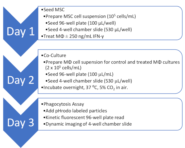
MSC and macrophage co-culture steps; diagram details 3-day protocol and phagocytosis assay.
Figure 1: Overview of the co-culture and phagocytosis assay workflow. An outline of the workflow for quantitative and qualitative analysis of MΦ phagocytosis activity in co-culture with MSC.

Supplementary File 1: A repres.......

Access restricted. Please log in or start a trial to view this content.

Disclosures

No conflicts of interest declared.

Materials

List of materials used in this article
NameCompanyCatalog NumberComments
96-well Black Polystyrene MicroplateMilliporeSigmaCLS3603-48EA
0.4% trypan blue solutionMilliporeSigmaT8154-20ML
15 mL and 50 mL Conical Sterile Polypropylene Centrifuge TubesThermoFisher339653
Antibiotic-Antimycotic (100X) GibcoThermoFisher15240096
Cell lifterMilliporeSigmaCLS3008-100EA
Culture flasks, tissue culture treated, surface area 75 cm2, canted neck, with cap, filteredMilliporeSigmaC7231-120EA
D1 ORL UVA [D1]ATCCCRL-12424Mouse MSC Cell Line
DMEM, High GlucoseThermoFisher11965092
Fetal Bovine Serum, qualified, heat-inactivatedThermoFisher16140071
HemocytometerFisherScientific02-671-51B
I-11.15ATCCCRL-2470Mouse MΦ Cell Line
LADMAC Cell LineATCCCRL-2420LADMAC cells secrete the growth factor colony stimulating factor 1 (CSF-1).
Live-Cell Imaging solutionThermoFisherA14291DJ
PBS, pH 7.4ThermoFisher10010031
pH Rhodo Green Zymosan Bioparticles ConjugateThermoFisherP35365
Spectramax i3XMolecular Devices
Sterile Single-Use Vacuum Filter Units, 250 mL, 0.2 µmThermoFisher568-0020
Sterile syringe filters, 0.2 micrometerThermoFisher723-2520
Tissue-culture-treated culture dishes, 100 mm x 20 mmMilliporeSigmaCLS430167-100EA
Trypsin-EDTA (0.05%), phenol redThermoFisher25300054

Tags

MSC-Macrophage Co-cultureZymosan ParticlespH-Sensitive FluorophoreTunneling NanotubesMitochondrial TransferFluorescence Intensity MeasurementLive-Cell ImagingPhagolysosome Formation