Method Article

Calcein Acetoxymethyl Staining to Evaluate Natural Killer Cell-Mediated Cytotoxicity toward Cancer Cells

1.2K views

July 8th, 2025

In This Article

Abstract

Source: Chava, S. et al., Measurement of Natural Killer Cell-Mediated Cytotoxicity and Migration in the Context of Hepatic Tumor Cells. J. Vis. Exp. (2020)

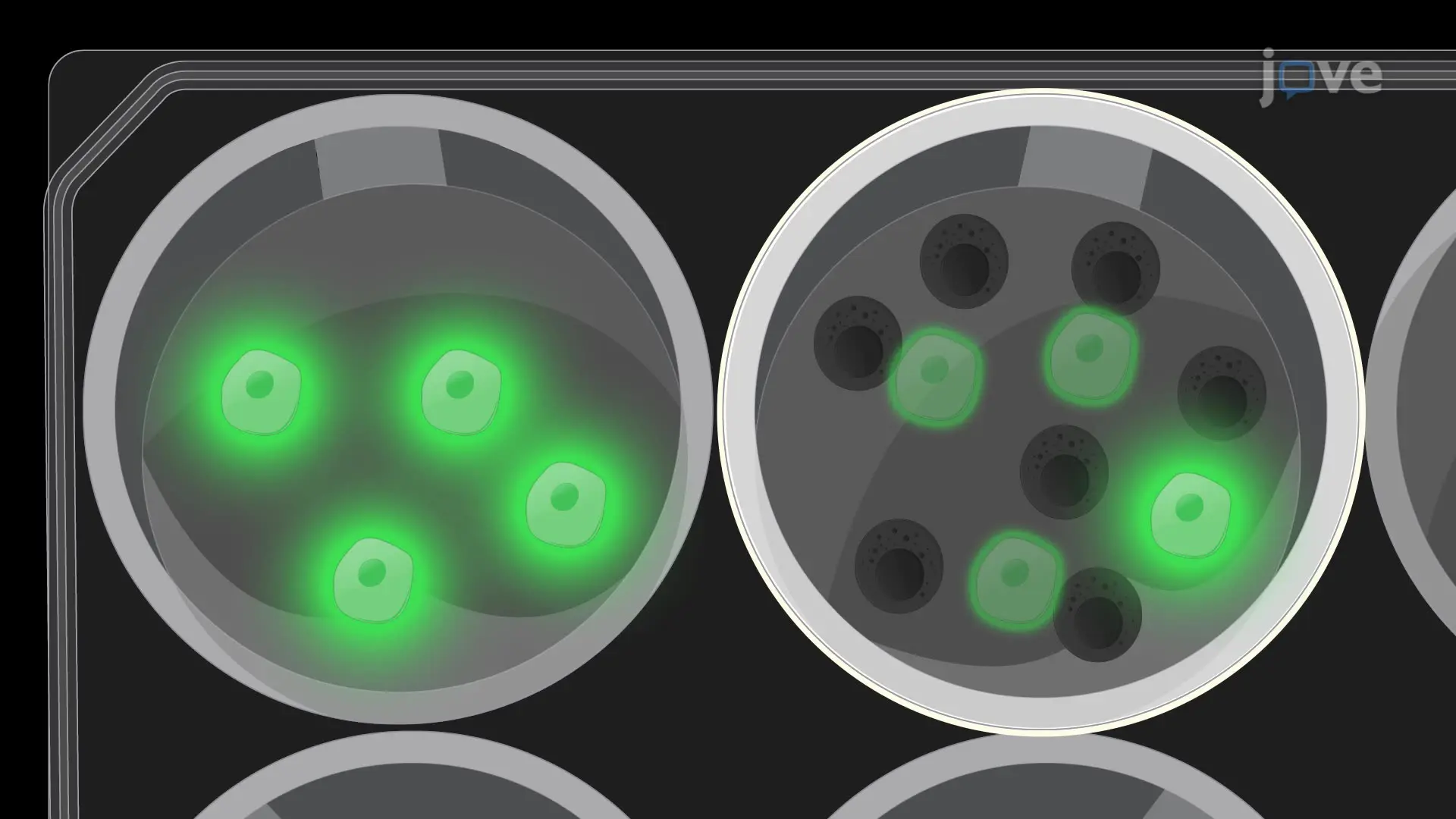
This video demonstrates an assay to measure the cytotoxicity of natural killer cells against cancer cells using the Calcein AM stain. The reduced number of fluorescent cancer cells following co-culture with natural killer cells indicates natural killer cell-mediated cytotoxicity toward cancer cells.

Protocol

1. Calcein AM Staining-based Microscopic Method for Measuring NK Cell-mediated Cytotoxicity

  1. Culture SK-HEP-1 cells to 70−80% confluency. Generate a single-cell suspension by first washing cells with 5 mL of 1x PBS followed by incubation with 1 mL of 0.25% trypsin-EDTA.
  2. Centrifuge SK-HEP-1 cells in a 1 mL sterile centrifuge tube at 160 x g for 3 min. Resuspend the pellet in 3 mL of serum-free DMEM.
  3. Add 1.5 µL of calcein AM solution (10 mM) to SK-HEP-1 cells and incubate for 30 min at room temperature. Centrifuge calcein AM-labeled SK-HEP-1 cells at 160 x g for 3 min in a 15 mL sterile centrifuge tube.
  4. Wash cells twice with 5 mL of 1x PBS to remove excess calcein AM dye.
  5. In parallel, centrifuge NK92MI cells at 160 x g for 3 min in a 15 mL sterile centrifuge tube. Wash the cell pellet once with 5 mL of 1x PBS and resuspend in 5 mL of NK92MI cell medium.
  6. Count calcein AM-labeled SK-HEP-1 cells and NK92MI cells using a hemocytometer or an automated cell counter.
  7. Resuspend SK-HEP-1 cells in the culture medium at 1 x 10⁵ cells/mL and NK92MI cells at 1 x 10⁶ cells/mL in the NK cell medium.
  8. Plate calcein AM-labeled SK-HEP-1 cells (target cells) (1 x 10⁴/100 µL/well) with NK92MI cells (effector cells) (1 x 10⁵/100 µL/well) (1:10 target: effector ratio) per well in triplicate wells in a 96 well plate.
  9. Incubate the 96-well plates at 37 °C in an atmosphere of 95% air and 5% CO2 for 4 h. After incubation, capture fluorescence images of the calcein AM-labeled cells using a fluorescence microscope at 10x magnification. Capture at least 10 different fields of each replicate for each treatment condition.
  10. Randomly select 10 images for each replicate and count calcein AM-positive labeled target cells incubated with or without NK92MI cells. Calculate % cytotoxicity using the formula below.
    NK cell cytotoxicity formula, diagram; ratio of target cell death with NK92MI, immune response calculation.
    NOTE: As controls, use target cells without NK92MI cells and completely lysed target cells as complete lysis control. For complete lysis, incubate the cells in 0.5% Triton X-100 for 1 h (20 µL of 5% Triton X-100 in 200 µL of culture media).

Access restricted. Please log in or start a trial to view this content.

Disclosures

No conflicts of interest declared.

Materials

List of materials used in this article
NameCompanyCatalog NumberComments
Calcein AM dyeSigma-Aldrich17783
DMEMGibco11965-092
Fetal Bovine SerumGibco26140079
Horse SerumGibco16050114
NK92MI cellsATCCCRL-2408
Phosphate Buffer Saline (PBS)Sigma-AldrichP4417
SKHEP-1 CellsATCCHTB-52
Trypsin EDTA solutionGibco25200056

Reprints and Permissions

Request permission to reuse the text or figures of this JoVE article

Request Permission

Tags

Calcein AM StainingNatural Killer Cell CytotoxicityCancer Cell AssayFluorescence MicroscopySerum Free MediaCytotoxicity MeasurementCell Co CultureApoptosis DetectionFlow CytometryFluorescence Imaging

Related Articles