All procedures involving human participants have been performed in compliance with the institutional, national, and international guidelines for human welfare and have been reviewed by the local institutional review board.
1. Preparation of materials
- Store reagents, antibodies, and chemicals as shown in the Material Safety Data Sheet (MSDS). Dissolve the drugs or cytokines in solvents as stock solutions and then aliquot for storage at -20 °C or -80 °C.
NOTE: Detailed information for material preparation is noted in the Table of Materials.
2. Peripheral blood mononuclear cell (PBMC) isolation
- Warm the density gradient solution (Table of Materials) to 18–20 °C before use. Invert the solution bottle several times to ensure thorough mixing.
- Collect 3−5 mL of human venous blood sample in a heparinized vial and mix well by gently inverting the tube several times.
- Prepare 4 mL of density gradient solution in a 15 mL sterile tube.
- Carefully layer 1 mL of the blood sample onto the density gradient solution.
- Centrifuge at 400 x g for 30 min at 18−20 °C (turn off the break).
- Carefully and immediately aspirate the buffy coat layer of mononuclear cells (about 1 mL) at once to avoid disturbing the layers to a sterile 15 mL tube using a 1 mL sterile pipette.
- Add at least 3 volumes (~3 mL) of phosphate-buffered saline (PBS) to the buffy coat in the centrifuge tube. Suspend the cells by gently pipetting them up and down at least 3x with a sterile pipette.
- Centrifuge at 400 x g for 10 min at 18−20 °C. Aspirate the supernatant.
- Suspend the cell pellet with 5 mL of basal medium (Table of Materials) and transfer into a flask. Culture the cells in a cell culture incubator at 37 °C and 5% CO2.
3. CIK induction and expansion
- On Day 0, culture the PBMCs (1 x 10⁶) in fresh basal medium containing 1,000 IU/mL of IFN-γ for 24 h in a humidified cell culture incubator at 37 °C and 5% CO2.
- On Day 1, refresh the medium with fresh basal medium containing 50 ng/mL of anti-CD3 antibody, 1 ng/mL of recombinant human (rh) IL-1α, and 1,000 U/mL of rh IL-2. Refresh the medium every 3 days.
- On Day 7, refresh the medium with fresh basal medium containing 1,000 U/mL of rh IL-2. Refresh the medium every 3 days until the end of cell expansion (Day 14).
4. Immunophenotyping for assessment of CIK cells
- Wash the CIK cells with 10 mL of sterile PBS. Centrifuge for 10 min at 300 x g and 18−20 °C, aspirate the supernatant, and resuspend the cells with 10 mL of PBS. Count the cell number and test cell viability using the trypan blue exclusion assay.
- Aliquot the CIK cells into six sterile 1.5 mL tubes at a density of ~5–10 x 105 cells/mL PBS. Label and treat as follows: Tube 1, Blank (no antibody); Tube 2, add 20 µL of isotype IgG1-FITC; Tube 3, add 20 µL of isotype IgG1-APC mAbs; Tube 4, add 20 µL of CD3-FITC; Tube 5, add 20 µL of CD56-APC mAbs; and Tube 6, add 20 µL of CD3-FITC and 20 µL of CD56-APC mAbs.
- Gently mix the CIK cells with the antibodies by gently pipetting them up and down at least 3x with a 1 mL sterile pipette, and then incubate for 30 min at room temperature in the dark.
- Centrifuge the tubes for 10 min at 300 x g and 18−20 °C. Aspirate the supernatant and suspend the cell pellet once with 1 mL of PBS. Gently pipet them up and down at least 3x with a 1 mL sterile pipette.
- Repeat step 4.4.
- Leave the tubes in the dark before flow cytometric analysis.
5. CD marker recognition
- Transfer the cell suspension to a sterile 5 mL polystyrene round bottom tube with a cell strainer cap (100 μm mesh) by gently pipetting through the cap. Put the tubes on the carousel in order.
- Open the flow cytometry analysis software and create an experimental folder. Click the New Specimen button to add a specimen and tube to the experiment and name the tubes as follows: Tube 1, Blank; Tube 2, Isotype IgG1-CD3; Tube 3, Isotype IgG1-CD56; Tube 4, CD3; Tube 5, CD56; Tube 6, CD3CD56.
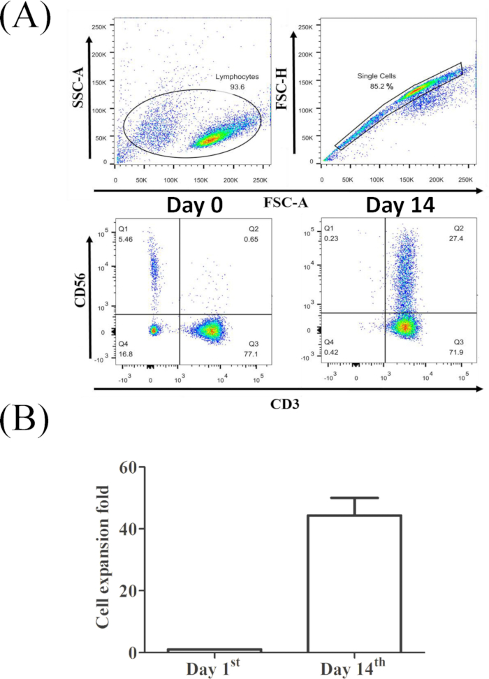
- Create a scatter gating system for the CIK cell populations (Figure 1A).
- Select Tube 1 (Blank) and click on the Dot Plot button to create an FSC-A/SSC-A plot. Draw a rectangle gate over the entire cell population with an FSC-A threshold >5 x 10⁴ to exclude cell debris.
- Select the SSC-A/SSC-H parameter for the new dot plot and draw a polygon gate around all single cells. Select the Count/FITC (CD3) and Count/APC (CD56) parameter for the new histogram plot, respectively. Select the FITC (CD3)/APC (CD56) parameter for the new dot plot and draw a four quadrant gate to define the four subpopulations.
- Record the data from 20,000 single cells in each specimen. Click the Load Sample button to analyze the Blank control sample first. Identify the whole CIK cell population by using the CD56 and CD3 channel parameters.
- Repeat step 5.3 for the investigation of all specimens.
- Open the files containing the statistical values of the individual specimen to analyze CIK cell populations and reprint them into analysis files.
6. Culturing and staining of human chronic myeloid leukemia K562 cells and ovarian cancer OC-3 cells
- K562 cells
- Culture K562 cells in complete media (RPMI basal medium containing 10% fetal bovine serum [FBS] and 50 U/mL antibiotics and adjust glucose to 4.5 g/L) at a density of 0.5−1 x 10⁶ cells/mL in a cell culture flask and incubate in a humidified incubator at 37 °C and 5% CO2.
- Transfer the culture media containing the K562 cells into 50 mL sterile tubes and pellet the cells at 300 x g for 10 min at 18−20 °C on the day of the experiment.
- Aspirate the supernatant, resuspend the cells in 5 mL of sterile PBS, and mix well gently.
- Pellet the cells at 300 x g for 10 min. Aspirate the supernatant, resuspend the cells in PBS, and adjust the K562 cells to a concentration of 0.5-1 x 10⁶ cells/mL.
- Add 0.5 µL of carboxyfluorescein succinimidyl ester (CFSE) dye to the 1 mL of K562 cell suspension in a 15 mL sterile tube at a final concentration of 5 μM. Gently mix the suspension by pipetting up and down at least 3x.
- Leave the tube in a cell culture incubator at 37 °C and 5% CO2 for 10–15 min.
- Add 9 mL of PBS to the tube and pellet the cells at 300 x g for 10 min. Decant the supernatant and then suspend the cell pellet in 10 mL of complete media. Transfer the cell suspension to a cell culture flask and place in the incubator.
- OC-3 cells
- Culture OC-3 cells in complete media (DMEM/F12 medium containing 10% FBS and 50 U/mL antibiotics) at a density of 0.5–1 x 10⁶ cells in a cell culture flask at 37 °C and 5% CO2.
- Aspirate the culture media and wash the cells with PBS 1 day before the experiment.
- Detach the cells by adding 1 mL of cell dissociation enzyme solution (Table of Materials) and incubate for 5 min at 37 °C.
- Suspend the cells by adding 5 mL of PBS and mix well gently. Pellet the cells at 300 x g for 10 min and aspirate the supernatant. Resuspend the cells in PBS and adjust the cells to a concentration of 0.5–1 x 10⁶ cells/mL.
- Add 0.5 µL of CFSE dye to 1 mL of the OC-3 cell suspension in a 15 mL sterile tube at a final concentration of 5 μM. Gently mix the suspension by pipetting up and down at least 3x.
- Leave the tube in a cell culture incubator at 37 °C and 5% CO2 for 10–15 min.
- Add 9 mL of PBS to the tube and pellet the cells at 300 x g for 10 min. Decant the supernatant and then suspend the cell pellet with complete media. Seed 5 x 10⁵ cells/well into a 6 well plate and incubate in a humidified incubator at 37 °C and 5% CO2 overnight.
7. Cytotoxic assay
- Coculture of CIK and K562 cells (CIK-K562)
- Count the K562 cells from step 6.1.7 and test the cell viability by trypan blue exclusion assay. Add 1 mL of K562 cells to each well in a 6 well plate at a density of 5 x 10⁵/mL.
- Add 1 mL of basal medium with or without CIK cells from step 3.4 to the 6 well plate from step 7.1.1 as follows: Well 1 = Blank, K562 cells alone (5 x 10⁵); Well 2 = CFSE-stained K562 cells alone (5 x 10⁵); Well 3 = CIK cells (E [effector], 25 x 105) + CFSE-stained K562 cells (T [target], 5 x 10⁵); Well 4 = CIK cells (E, 50 x 105) + CFSE-stained K562 cells (T, 5 x 10⁵).
- Mix the cell suspensions by gently pipetting them up and down at least 3x. Place the plate in the incubator for 24 h.
- Coculture of CIK and OC-3 cells (CIK-OC-3)
- Add 1 mL of basal medium with or without CIK cells from step 3.4 to the 6 well plate from step 6.2.7 as follows: Well 1 = Blank, OC-3 cells alone (5 x 10⁵); Well 2 = CFSE-stained OC-3 cells alone (5 x 10⁵); Well 3 = CIK cells (E, 25 x 105) + CFSE-stained OC-3 cells (T, 5 x 10⁵); Well 4 = CIK cells (E, 50 x 105) + CFSE-stained OC-3 cells (T, 5 x 10⁵).
- Mix the cell suspensions by gently pipetting them up and down at least 3x. Put the plate in the incubator for 24 h.
- 7-Aminoactinomycin D (7-AAD) dye staining
- Harvest the CIK-K562 cell suspension from step 7.1.3 directly into a 15 mL sterile tube.
- Harvest both the suspension and adherent cells from the CIK-OC-3 groups from step 7.2.2.
- Transfer the cell suspension to a 15 mL sterile tube. Wash the well with 1 mL of sterile PBS, collect the PBS, and add to the tube. Add 0.5 mL of cell dissociation enzyme solution, and incubate for 5 min at 37 °C.
- Add 1 mL of the solution from the same tube to the corresponding well and gently mix the cells by pipetting them up and down at least 3x with a 1 mL sterile pipette. Collect all the cells in the same tube.
- Centrifuge at 300 x g for 10 min, aspirate the supernatant, and resuspend the cells in 1 mL of sterile PBS. Pellet the cells at 300 x g for 10 min, aspirate the supernatant, and resuspend cells in 100 µL of sterile PBS.
- Add 5 µL of 7-AAD dye (50 ng/µL stock) to the cell suspension. Gently mix the cells by pipetting them up and down at least 3x with a 1 mL sterile pipette. Incubate for 10 min and leave in the dark before analysis.
- Cytolytic capability assay
- Mix the cell suspension from step 7.3.4 and repeat steps 5.1 and 5.2 once.
- Click the New Specimen button to add a specimen and tube to the experiment and name the tubes as follows: Tube 1, K562 (or OC-3) cells only; Tube 2, CFSE-stained K562 (or OC-3) cells only; Tube 3, E:T = 5:1; Tube 4, E:T = 10:1.
- Create a Scatter Gating System for the cytolytic assay (Figure 2A).
- Select Tube 1 and click on the Dot Plot button to create an FSC-A/SSC-A plot. Draw a rectangle gate over all events with an FSC-A threshold >5 x 10⁴ to exclude cell debris.
- Select the SSC-A/CFSE parameter for the new dot plot. Select the 7-AAD/CFSE parameter for the new dot plot and draw a four-quadrant gate to define the four subpopulations.
- Click the Load Sample button to analyze the blank control sample first.
- Adjust the voltage of SSC-A and FSC-A. Identify the dead cell population by using the CFSE and 7-AAD channel parameters. Record the data from >20,000 CFSE+ cells in each specimen.
- Repeat section 7.4.6 for the investigation of all specimens.
- Open the files containing the statistical values of each individual specimen to analyze the non-viable cell populations and export the data into analysis files.