A subscription to JoVE is required to view this content. Sign in or start your free trial.

Method Article

Photoconversion-Based Leukocyte Tracking in Bacterial-Infected Transgenic Zebrafish

856 views

⸱

July 8th, 2025

In This Article

Abstract

Source: Harvie, E. A. et al., Non-invasive Imaging of the Innate Immune Response in a Zebrafish Larval Model of Streptococcus iniae Infection. J. Vis. Exp. (2015)

This video demonstrates the tracking of leukocyte recruitment using transgenic zebrafish larvae expressing photoconvertible green fluorescent protein. Pre-injecting red fluorescent dye-labeled bacteria into the otic vesicle and inducing photoconversion enables observation of leukocytes' migration, phagocytosis, and dispersal throughout the body, providing valuable insights into immune response dynamics.
Protocol

1. Preparing Microinjection Needles

  1. Prepare thin wall glass capillary injection needles (1.0 OD/0.75 ID) using a micropipette puller device with the following settings: air pressure 200, heat 502, pull 90, velocity 80, time 70, air time at the start of pull 5, air time at end of pull 5.
  2. Using fine tweezers, break off the tip of the pulled needle so that the tip opening has a diameter of approximately 10 µm.

2. Preparing Larval Injection Dishes

  1. Rinse a gel comb with a lane width of approximately 4-5 mm in sterile water and allow it to dry.
  2. Prepare a 1.5....

Access restricted. Please log in or start a trial to view this content.

Results

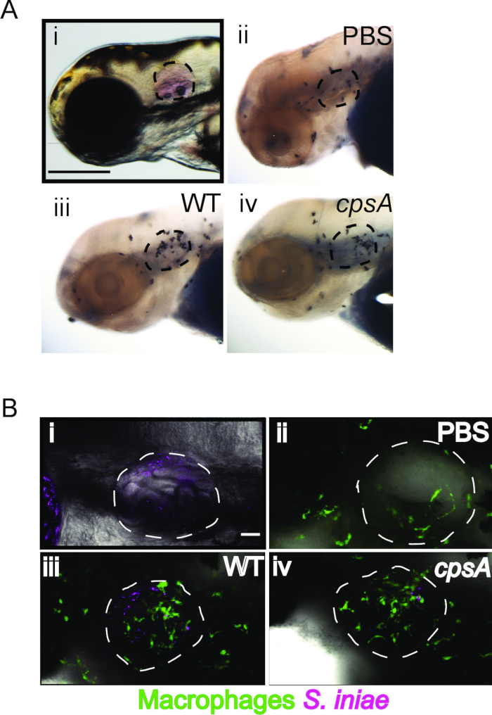
Microscope images of macrophage interactions with S. iniae; immunohistochemistry analysis diagram.
Figure 1: Leukocyte recruitment to otic vesicle infection with S. iniae. (A) Neutrophil recruitment to S. iniae infection. (i) Successful injection of a phenol red-labeled inoculum into the otic vesicle........

Access restricted. Please log in or start a trial to view this content.

Disclosures

No conflicts of interest declared.

Materials

List of materials used in this article
NameCompanyCatalog NumberComments
1.7 ml eppendorfsMidSciAVSS1700
14 ml falcon tubeBD Falcon352059
96 well plateCorning Incorporated3596
AgarBD Biosciences214030
CellTracker RedMolecular Probes, InvitrogenC34552
CNA agarDot Scientific, Inc7126A
Disposable transfer pipetsFisher Scientific13-711-7m
Dissecting ScopeNikonSMZ745
DMSOSigma AldrichD2650
Fine tweezersFine Science Tools11251-20
Gel combVWR27372-4824.2 mm width, 1.5 mm thick
Glass bottom dishesCustom made by drilling a 16–18 mm hole in the center of a 35-mm tissue culture dish bottom and placing a 22-mm round #1 coverslip in the hole and sealing with a thin layer of Norland Optical Adhesive 68 cured by UV light.
GlycerolFisher ScientificG33-4
High melt agaroseDenville Scientific, Inc.CA3510-6
Laser Scanning Confocal MicroscopeOlympuswith FV-1000 system
Low melt agaroseFisherBP165-25
Magnetic standTritech (Narishige)GJ-1
Microinjection systemParkerPicospritzer III
Microloader pipet tipsEppendorf930001007
MicromanipulatorTritech (Narishige)M-152
Micropipette pullerSutter Instrument CompanyFlaming/Brown P-97
Nanodrop spectrophotmeterThermo ScientificND-1000
N-Phenylthiourea (PTU)Sigma aldrichP7629
ParaformaldheydeElectron Microscopy Sciences15710
Petri DishesFisher ScientificFB0875712100 mm x 15 mm
Phenol RedRicca Chemoical Company572516
Phosphate Buffered SalineFisher ScientificBP665-1
PronaseRoche165921
Protease peptoneFluka Biochemika29185
Small cell culture dishCorning Incorporated43016535 mm x 10 mm
Thin wall glass capillary injection needlesWorld Precision Instruments, Inc.TW100-3
Todd HewittSigma Aldrich/Fluka AnalyticalT1438
Tricaine (ethyl 3-aminobenzoate)Argent Chemical Laboratory/FinquelC-FINQ-UE-100G
Yeast extractFluka Biochemika92144

Explore More Articles

Photoconversion-Based TrackingLeukocyte RecruitmentBacterial Infection ModelFluorescent Protein PhotoconversionOtic Vesicle ImagingZ-Stack Scanning405-Nanometer LaserFluorescence MicroscopyImmune Cell Migration