A subscription to JoVE is required to view this content. Sign in or start your free trial.

Method Article

An Immunofluorescence Assay to Characterize and Quantify Mammalian Stress Granules

901 views

July 8th, 2025

In This Article

Abstract

Source: Aulas, A. et al., Methods to Classify Cytoplasmic Foci as Mammalian Stress Granules. J. Vis. Exp. (2017)

This video demonstrates an assay for characterizing and quantifying mammalian stress granules, which consist of mRNAs, RNA-binding proteins (RBPs), and various other proteins.

Protocol

1. Cell Preparation

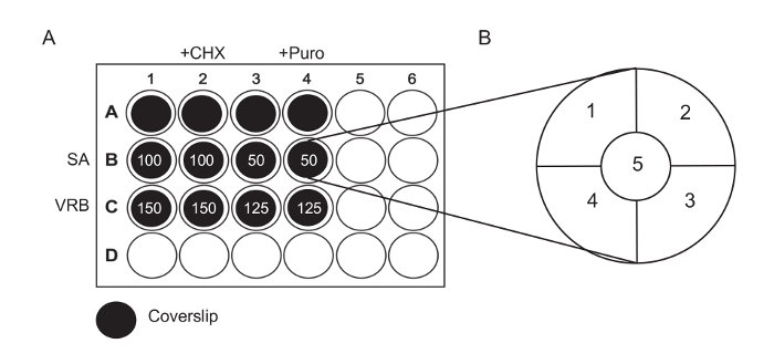
  1. Add autoclaved coverslips to 12 wells of a 24-well plate, as indicated in Figure 1A; this can be done using a sterile Pasteur pipette attached to a vacuum to pick up each coverslip. Gently tap the coverslip on the side of the well to release the suction, allowing the coverslip to fall into the well.
  2. Plate 1 x 10⁵ U-2 OS (U2OS) osteosarcoma cells per well at a final volume of 500 µL of medium. Move the plate side to side and up and down several times to ensure that the cells are evenly distributed.
  3. Gently press the coverslips down with a clean P1000 tip to ensure that the coverslip is on the bottom of the well and that there are no bubbles under the coverslip.

2. Stressing Cells

  1. The following day, visually check the plate to ensure that the cells are evenly dispersed and evenly confluent across the coverslip, as variations between samples may adversely impact the reproducibility of results. Use a 10X objective on an inverted tissue-culture microscope.
  2. Preheat the medium to 37 °C. Avoid applying cold or hot media to the cells, as these cause cold shock or heat shock, respectively.
  3. Dilute drugs in preheated media. For each different drug concentration, prepare 2 wells containing 500 µL of medium. Prepare an additional 500 µL to allow for loss while pipetting. Aliquot 4 tubes, each containing 1.5 mL.
    1. For sodium arsenite: to each well containing 500 µL, add 1.5 µL of 100 mM sodium arsenite (final concentration: 100 µM) or add 0.75 µL of 100 mM sodium arsenite (final concentration: 50 µM).
    2. For vinorelbine: to each well containing 500 µL, add 22.5 µL of 10 mM vinorelbine (final concentration: 150 µM) or add 18.75 µL of 10 mM vinorelbine (final concentration: 100 µM).
      NOTE: Sodium arsenite is a well-characterized and commonly used stress granule inducer and is therefore classically used as a positive control.
  4. Remove and discard the media from wells B1 - 4 and C1 - 4. Add the media with drugs and wait for 60 minutes (Figure 1A).
    1. Add 500 µL of medium with 100 µM sodium arsenite to wells B1 and B2. Add 500 µL of medium with 50 µM sodium arsenite to wells B3 and B4.
    2. Add 500 µL of medium with 150 µM vinorelbine to wells C1 and C2. Add 500 µL of medium with 125 µM vinorelbine to wells C3 and C4.
  5. 30 min prior to fixation, treat the wells in column 2 with 5 µL of cycloheximide (1 mg/mL stock) and the wells in column 4 with 2 µL of puromycin (1.25 mg/mL stock). Gently rock the plate to mix before placing the plate back in the 37 °C incubator.

3. Cell Fixation and Immunofluorescence

NOTE: Before the experiment, ensure that the methanol is cooled to -20 °C. Make buffers, including Phosphate-buffered Saline (PBS), 5% Normal Horse Serum (NHS) in PBS, and 4% paraformaldehyde (PFA) in PBS.

  1. Discard the media and wash the wells containing coverslips with PBS. Depending on the drugs used, it might be important to properly discard the media-containing drugs; contact the local environmental safety office for specific instructions. To wash efficiently, fill a squeeze bottle with PBS, cut back the tip in order to allow a gentle flow rather than a tight stream, and use this to add PBS to each well after aspirating the original PBS.
  2. Fix the cells using ~250 µL of 4% PFA for 15 min at room temperature, RT under gentle agitation. Completely cover the top of the coverslip with PFA; 250 µL should be sufficient, but if not, add more. Always avoid pipetting directly onto the coverslips, as some cells (or stress treatments of cells) can alter attachment, and the force from pipetting can dislodge the cells.
  3. Remove the PFA and discard properly. Contact the local environmental safety office for details on how to properly discard paraformaldehyde.
  4. Add ~250 µL of methanol (-20 °C) and incubate for 5 min at RT under gentle rocking; this both permeabilizes and flattens the cells.
  5. Remove and discard the methanol and block the cells by applying ~250 µL of 5% NHS for 1 h at RT overnight, O/N, at 4 °C. Contact the local environmental safety office for details on how to properly discard methanol.
  6. For primary antibodies, dilute the antibodies in 5% NHS.
    NOTE: The use of multiple SG markers is key to confirming whether the granules observed are bona fide SGs. The number of SG markers that can be used depends on the filters available in the microscope. If standard green, red, and far-red filter sets are available, use G3BP1, eIF4G, and eIF3b at a 1/250 dilution.
    1. For this experiment (12 coverslips at 250 µL each-Total: 3 mL of 5% NHS), add 12 µL of each of the following: anti-G3BP1, anti-eIF3b, and anti-eIF4G. Incubate the primary antibodies using gentle agitation for at least 1 h at RT or O/N at 4 °C.
  7. Wash the wells 3x with PBS, and incubate each wash for 5 min.
  8. For secondary antibodies, dilute all secondary antibodies (1/250 dilution) and Hoechst dye (1/1,000 dilution) in 5% NHS.
    1. For this experiment (12 coverslips at 250 µL each-Total: 3 mL of 5% NHS), add 12 µL of each of the following: Cy2-Mouse, Cy3-Rabbit, and Cy5-Goat (a 1/250 dilution of each) and add 3 µL of Hoechst dye.
    2. Incubate the coverslips with the secondary antibodies for 1 h at RT. During this and the following steps, protect the samples from light by covering the plate with a box or by placing foil over the top of the tissue culture dish.
  9. Wash the wells three times with PBS for 5 min each.
  10. Mount the coverslips onto labeled glass slides using a mounting medium. Heat the mounting medium in a 37 °C heat block for 10 min; this decreases the viscosity and makes it easier to pipette. With a cut P200 tip, pipette 25 µL of mounting medium per coverslip onto a glass slide; 4-8 coverslips can fit on one glass slide, assuming that the microscope stage allows this. Using fine forceps, transfer the coverslip from the well to the slide, making sure to put the cell side down on the mounting medium. Using a clean P200 tip, press the coverslip down.
  11. Once all the coverslips have been mounted, use folded lab tissue to clean up the excess mounting medium by pressing the lab tissue very firmly onto the slide. Use a squeeze bottle containing H2O to flush off the excess mounting medium and then repeat the blotting with folded lab tissue to remove the water.

Access restricted. Please log in or start a trial to view this content.

Results

Multiwell plate analysis diagram, cycloheximide/puro treatments, cellular response quantification.
Figure 1: Experimental Diagrams. (A) Seed cells onto coverslips previously placed in a 24-well plate, as indicated. Treat Rows A and B for 60 min with SA or VRB using the concentration in µM as indicated. After 60 min, add CHX (final...

Access restricted. Please log in or start a trial to view this content.

Disclosures

No conflicts of interest declared.

Materials

List of materials used in this article
NameCompanyCatalog NumberComments
U-2 OSATCCATCC®HTB-96™- commonly written as "U2OS"
- culture at 37 °C under 5% CO2 in 10% FBS/DMEM
Dulbecco's Modification of Eagle's MediumCorning10-013-CV- abbreviated DMEM
- contains 4.5 g/L glucose, pyruvate, and phenol red
- supplemented with 10% FBS, 10 mM HEPES, and penicillin/steptomycin
- prewarm prior drug(s) dilution
Fetal Bovine SerumSigmaF2442-500ml- abbreviated FBS
- used to supplement media
HEPES (1 M)Thermo Fisher Scientific15630-080- used to supplement media
Penicilline StreptomycineSigmaP0781-100ml- used to supplement media
24 well plateCostar3524
Lab tissuesKIMTECH SCIENCE34120- commonly called "kimwipes"
Coverglass for Growth Cover GlassesFisher Scientific12-545-82- autoclaved before used
- keep sterile in the tissue culture hood
Sodium (meta)arsenite ≥90%SigmaS7400-100G- commonly called sodium arsenite and abbreviated SA
- dissolution in water at 1 M concentration, then dilute to 100 µM as a working stock
VinorelbineTSZ CHEMRS055- abbreviated VRB
- dissolve in water to 10 mM
PuromycinSigmaP9620- used to assess translation level (ribopuromycylation)
- used to assess SG connection with active translation
- dilute in water to 10 mg/mL
Emetine dihydrochloride hydrateSigmaE2375- used in combination with puro to assess general translation level
- used to assess SG connection with active translation
- dilute in water to 10 mg/mL
CycloheximideSigmaC4859- abbreviated CHX
- used to assess SG connection with active translation
- dilute in water to 10 mg/mL
Phosphate Buffered SalineLonza / VWR95042-486- abbreviated PBS
- wash buffer for immunofluorescence
Paraformaldehyde reagent grade, crystallineSigmaP6148-500G- make a 4% solution in hot PBS in fume hood, stir until dissolved
- aliquots can be stored at - 20 °C for several months
- hazardous, use ventilation
- discard in special waste
Methanol, ACSBDH / VWRBDH1135-4LP- pre-chilled to -20°C before use
- discard in special waste
Normal Horse SerumThermo Fisher Scientific31874- abbreviated NHS
- dilute to 5% in PBS
- add sodium azide for storage at 4°C
- blocking solution for immunofluorescence
Ethanol, Pure, 200 Proof (100%),USP, KOPTECDecon Labs / VWR89125-188- dilute to 70% with water
Fisherbrand Superfrost Plus Microscope SlidesFisherbrand12-550-15
Mouse anti G3BP1 antibody (TT-Y)Santa Cruzsc-81940- store at 4 °C
- use at 1/100 dilution
Rabbit anti eIF4G antibody (H-300)Santa Cruzsc-11373- store at 4 °C
- 1/250 dilution
Goat anti eIF3η (N-20) antibodySanta Cruzsc-16377- eIF3η is also known as eIF3b
- store at 4 °C
- 1/250 dilution
Cy2 AffiniPure Donkey Anti-Mouse IgG (H+L)Jackson Immunoresearch715-225-150- reconstitute in water as per manufacturer's instructions then store at 4°C
- 1/250 dilution
Cy3 AffiniPure Donkey Anti-Rabbit IgG (H+L)Jackson Immunoresearch711-165-152- reconstitute in water as per manufacturer's instructions then store at 4°C
- 1/2500 dilution
Cy5 AffiniPure Donkey Anti-Goat IgG (H+L)Jackson Immunoresearch705-175-147- reconstitute in water as per manufacturer's instructions then store at 4°C
- 1/250 dilution
Hoechst 33258 solutionSigma94403-1ML- incubate with secondary antibodies-stock solution 0.5 mg/mL in dH20, protect from light- Store at 4 °C
20 x SSC bufferThermo fisher AmbionAM9763- dilute to 2X SSC using RNase Free water (DEPC treated)- store at room temperature- wash buffer for FISH
Slide mounting mediahome made/- mix 5 g of "cold-soluble" poly(vinyl alcohol) in 20 ml of PBS- mix by sonication, followed by stirring overnight at RT- Add 5 ml of glycerol and 0.2 ml of 20 % sodium azide, and stir for 16 h at RT- centrifugation at 20,000 g for 20 min, discard large pellet- aliquot viscous liquid, long-term storage at -20°C, 1 week at 4°C
Parafilm "M"SigmaP7793-1EA- usually referred as "parafilm"
Poly(vinyl alcohol)SigmaP-8136-250G- reagent to make vinol
GlycerolSigmaG5516-100ML- reagent to make vinol
Sodium azideFisher ScientificS2002-5G- preservative agent for blocking solution and vinol
- make a 20 % dilution in water

Tags

Fluorescence MicroscopyPrimary AntibodiesSecondary AntibodiesHoechst DyeCell FixationCell PermeabilizationBlocking BufferMounting Medium