1. Preparation of target cells that express red fluorescent protein in their nuclei
- Obtain a target mouse cancer cell line from an appropriate source.
NOTE: In this protocol, a highly metastatic derivative of E0771 mouse mammary tumor cells (E0771-LG) is used. Parental E0771 cells originate from C57BL/6 mice.
- Thaw and maintain a vial of E0771-LG cells with Dulbecco's Modified Eagles Medium (DMEM) including 10% (v/v) fetal bovine serum (FBS) in a cell culture incubator at 37 °C, 95% humidity, and 5% CO2.
NOTE: It should be confirmed that cells are negative for mycoplasma. To this end, culture E0771 cells (or cells to be tested) for 2-3 days as described above (in the absence of antibiotics and antimycotics), and collect 500 μL of culture medium. Centrifuge the medium at 12,419 x g for 60 s to eliminate cell debris and transfer the supernatant into new tubes. Determine mycoplasma contamination using a commercially available mycoplasma test kit (refer to Table of Materials) and/or PCR following the manufacturer's instructions.
- Seed 5 x 10³ E0771-LG cells per well into a 12-well plate, and culture the cells with 10% (v/v) FBS-DMEM overnight in an incubator at 37 °C, 95% humidity, and 5% CO2.
NOTE: If the proliferation rate of target cells is low (population doubling time greater than 36 h), the number of cells can be increased to 1 x 10⁴.
- Replace the medium with 1 mL of 10% (v/v) FBS-DMEM including 10 μg/mL polybrene and add 25 μL of lentiviral particles (1 x 10⁶ TU/mL) encoding a nuclear restricted red fluorescent protein (mKate2, refer to Table of Materials).
- Culture the cells for 24 h in an incubator at 37 °C, 95% humidity, and 5% CO2.
- Replace the medium with 10% (v/v) FBS-DMEM and culture the cells for 24-48 h in an incubator at 37 °C, 95% humidity, and 5% CO2.
- Replace the medium with 10% (v/v) FBS-DMEM including 1 μg/mL puromycin when cells start to express a red fluorescent protein, and culture the cells until they are 80-90% confluent.
NOTE: The concentration of puromycin will be different between target cell types and should be optimized using un-transfected cells.
- Subculture the surviving cells for 1-3 passages with 10% (v/v) FBS-DMEM including 1 μg/mL puromycin, and cryopreserve stocks in a liquid nitrogen vapor phase storage system until use.
2. Isolation of suppressor cells from the tumors in mice
NOTE: In this protocol, suppressor cells (i.e., metastasis-associated macrophages (MAMs) and monocytic-myeloid-derived suppressor cells (M-MDSCs)) are isolated from the lung containing metastatic tumors established by E0771-LG cells. Conditions for tissue dissociation and cell sorting should be optimized to isolate the cells from different tissues.
- Inject 1 x 10⁶ cancer cells (E0771-LG) into the tail vein of syngeneic (C57BL/6), female, 7-10 week old mice.
- After 14 days, isolate the lung containing metastatic tumors and prepare single-cell suspensions from the perfused lungs via enzymatic digestion as previously described.
NOTE: In this protocol, four mice are injected with cancer cells and their metastatic lungs are combined to obtain sufficient suppressor cells.
- Incubate the single cell suspensions with anti-mouse CD16/CD32 antibody for 30 min on ice, and stained with fluorescent antibodies to CD45, F4/80, CD11b, Ly6C, and Ly6G (refer to Table of Materials) for another 30 min.
- Wash the stained cells once with 1 mL of PBS containing 2% (w/v) bovine serum albumin (BSA), and re-suspend the cell pellet with 500–1000 μL of PBS containing 2% (w/v) BSA.
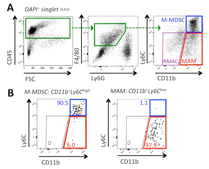
- Add 3 μM of DAPI, and sort M-MDSCs (DAPI–CD45+F4/80+Ly6G–CD11bhighLy6Chigh) and MAMs (DAPI–CD45+F4/80+Ly6G–CD11bhighLy6Clow) using a cell sorter (Figure 1).
NOTE: The threshold of Ly6C level to distinguish MAMs (Ly6Clow) and M-MDSCs (Ly6Chigh) is based on that of resident alveolar macrophages (RMAC). The purity of the sorted cells is measured via flow cytometry with an expected purity of more than 90%.
- Resuspend the sorted cells with 400 μL of DMEM containing 20% (v/v) FBS, 1% (v/v) penicillin/streptomycin, 2 mM L-glutamine, 1% (v/v) non-essential amino acid, 1 mM sodium pyruvate, and 50 nM 2-mercaptoethanol (called enriched-DMEM, E-DMEM).
- Count the number of live cells using the Trypan blue exclusion method and adjust to 2 x 10⁶ cells/mL with E-DMEM.
- Keep the cells on ice until use.
3. Isolation of CD8+ T cells from the spleen of mice
- Isolate the spleen from a mouse that is syngeneic to the target cancer cell line (i.e., C57BL/6 mice in this protocol) as follows:
- Euthanize the animal by CO2 inhalation.
- Place the animal on a clean dissection board and wipe the skin with 70% (v/v) ethanol.
- Cut the abdominal skin using scissors to expose the spleen
- Isolate the spleen, which is located inferior to the stomach, and place it into a tube containing 5 mL of ice-cold PBS.
- Using the inner plunger of a sterile 5 mL syringe, grind the spleen on a 100 μm cell strainer set on a 50 mL tube.
- Pass the cells through the filter using a total of 10 mL of PBS.
- Centrifuge the cell suspension at 337 x g for 5 min, and aspirate the supernatant.
- Resuspend the cell pellet in 1 mL of PBS containing 2 mM ethylenediaminetetraacetic acid (EDTA) and 0.5% (w/v) BSA (running buffer) and filter through a 40 μm cell strainer.
- Count the live cell number and adjust to 1 x 10⁸ cells/mL using the running buffer.
NOTE: Keep a small aliquot of cells as a pre-enrichment sample for a purity check.
- Enrich for CD8+ T cells using a negative selection kit and a magnetic sorter (refer to Table of Materials).
- Transfer 1 x 10⁸ (1 mL) of the splenocyte cells to a 5 mL polystyrene round-bottom tube.
- Add 50 μL of biotinylated antibodies, and incubate at room temperature for 10 min.
- Add 125 μL of streptavidin-conjugated magnetic beads, and incubate at room temperature for 5 min.
- Add 1.325 mL of running buffer, and gently mix by pipetting.
- Place the tube into the magnet, and incubate at room temperature for 2.5 min.
- Pick up the magnet, and pour the enriched cell suspension into a new tube.
- Resuspend the enriched cells with 200 μL of E-DMEM (i.e., DMEM containing 20% (v/v) FBS, 1% (v/v) penicillin/streptomycin, 2 mM L-glutamine, 1% (v/v) non-essential amino acid, 1 mM sodium pyruvate, and 50 nM 2-mercaptoethanol).
- Count the number of live cells and adjust to 2 x 10⁶ cells/mL with E-DMEM. Keep the cells at 37 °C in a CO2 incubator until use.
NOTE: Keep a small aliquot of cells as a post-enrichment sample for a purity check.
- Determine the purity of CD8+ T cells by flow cytometry as follows:
- Take 1 x 10⁴ cells from pre-enrichment (step 3.6) or post-enrichment (step 3.9) samples and adjust the total volume of each to 100 μL using a running buffer.
- Incubate the single cell suspensions with anti-mouse CD16/CD32 antibody for 30 min on ice, and stain with fluorescent antibodies to CD45, CD3, CD4, and CD8 (refer to Table of Materials) for another 30 min.
- Wash the stained cells with 500 μL of PBS containing 2% (w/v) BSA, and re-suspend the cell pellet with 500–1000 μL of PBS containing 2% (w/v) BSA.
- Add 3 μM of 4',6-diamidino-2-phenylindole (DAPI), and determine the percentage of CD3+CD4–CD8+ cells in the total CD45+ cell population.
4. Activation and expansion of the isolated CD8+ T cells
- Aliquot 1 x 10⁵ cells per 50 μL CD8+ T cells (prepared in step 3.9) into wells of a U-bottom 96-well plate.
- Add 1 x 10⁵ cells per 50 μL suppressor cells (prepared in step 2.7) or 50 μL of E-DMEM into the wells.
- Prepare activation medium that consists of E-DMEM, 4 x 10⁴ U/mL colony-stimulating factor 1 (CSF-1), 240 U/mL interleukin-2 (IL-2), 8 μg/mL anti-mouse CD3ε antibody, and 16 μg/mL anti-mouse CD28 antibody.
NOTE: CSF-1 is not required for T cell activation, but is essential for survival of suppressor cells in this protocol (i.e., MAMs and M-MDSCs). Thus, it is retained in the co-culture of T cells with target cancer cells to maintain consistency in culture conditions. Since CSF-1 is found in nano-molar concentrations in all tissues and is required for monocyte/macrophage viability in vivo, this is a physiological context for these cells.
- Add 50 μL of activation medium (step 4.3) and 50 μL of E-DMEM with or without reagents to be tested.
- Place the plate into an incubator at 37 °C, 95% humidity, and 5% CO2, and culture the cells for 4 days.
5. Setup of co-culture of target cells with pre-activated CD8+ T cells
- Add 30 μL of 1:100 diluted growth factor-reduced soluble basement membrane matrix (Table of Materials) into the wells of flat bottom 96-well plate suitable for microscopy, and incubate at 37 °C in a CO2 incubator for at least 1 h.
- Prepare target cells (i.e., E0771-LG cells expressing nuclear-restricted red fluorescent protein).
- Incubate the target cells with 1 mL of 0.05% trypsin/EDTA at room temperature for 1 min, and harvest the cells by gentle pipetting.
- Add 9 mL of DMEM including 10% (v/v) FBS, and centrifuge the cell suspension at 337 x g for 5 min.
- Re-suspend the cells with 500 μL of E-DMEM, and count the number of live cells.
NOTE: Before counting the target cells, filter the cells through a 40μm cell strainer to produce a single-cell suspension.
- Adjust the density to 2 x 10⁴ cells/mL (= 1 x 10³ cells per 50 μL) by adding E-DMEM, and keep the cells on ice.
- Prepare effector cells (i.e., pre-activated CD8+ T cells).
- Resuspend the cells in a well of a 96-well plate (step 4.5) thoroughly by pipetting, and transfer the floating CD8+ T cells into a new 1.5 mL tube.
NOTE: MAMs and M-MDSCs tightly adhere to the well and are not detached by the pipetting.
- To collect the remaining cells, add 200 μL of PBS into the well and transfer it into the tube in step 5.3.1.
- Wash the cells with 1 mL of E-DMEM once (centrifuge at 337 x g) for 5 min, aspirate and discard supernatant), and resuspend them with 100 μL of E-DMEM.
- Count the number of live cells (using the trypan blue exclusion method), adjust the density to 1.6 x 10⁵ cells/mL (= 4 x 10³ cells/25 μL), and keep the cells on ice.
- Aspirate the basement membrane matrix from each well of the plate in step 5.1.
- Add 1 x 10³ cells/50 μL of target cells (step 5.2.4) into each well and mix well.
NOTE: To avoid edge effects due to evaporation of the medium, only the inner 60 wells of the 96-well plate should be used for analysis.
- Add 25 μL of E-DMEM including 4 x 10³ U/mL IL-2 and 10 μM fluorogenic caspase-3 substrate (refer to Table of Materials).
- Add 4 x 10³ cells/25 μL of CD8+ T cells (step 5.3.4) into appropriate wells and mix well.
NOTE: The presence of too many cells in a well makes the analysis more difficult. In this model, a 4:1 effector: target ratio (total cell number 5 x 10³ cells/well) was optimal, but an 8:1 ratio was suboptimal. Wells containing the following four controls are necessary to aid with data analysis: target cells at the density used for the co-culture wells (1 x 10³ cells/well) in medium with and without caspase-3 substrate, effector cells (1 x 10³ cells/well) in medium with and without caspase-3 substrate.
- Add 200 μL of PBS or sterile water into all empty wells (particularly wells on the periphery of the plate) to reduce evaporation of medium from experimental wells.
- Set the plate into a time-lapse fluorescence microscope that is maintained at 37 °C, 95% humidity, and 5% CO2.
NOTE: Shake the plate in a cross-pattern to distribute all cells evenly, and then allow the plate to remain at room temperature on a flat surface for 10-20 min before transferring to the incubator.
6. Imaging of the cells
NOTE: Detailed image acquisition settings will vary with the microscope and fluorophores used; the following general acquisition parameters should be employed for optimal results.
- Using an appropriate autofocus routine on the microscope, acquire images covering at least 25% of the total surface area in each experimental well of the 96-well plate.
- Set the microscope to acquire images in phase contrast as well as a fluorescent channel suitable for the nuclear-restricted red fluorescent protein (mKate2) and a fluorescent channel suitable for the green fluorogenic activated caspase-3 substrate (with excitation at 488 nm) (Figure 2).
- Capture images in experimental wells in phase contrast and the 2 fluorescent channels every 1 to 3 h for at least 72 h.