1. Protein density calibrations on BSLBs (Bead-supported lipid bilayers)
- Before taking the 5.00 ±0.05 µm diameter non-functionalized silica beads, mix the stock solution well and resuspend any big clumps of beads sedimented on the bottom of the flasks.
NOTE: Silica beads tend to sediment quickly, which might lead to counting errors. Mix vigorously by pipetting up and down half of the maximum volume of a P1000 micropipette.
- Dilute 1 µL of bead solution in 1,000 µL of PBS, count the beads using a hemocytometer chamber, and calculate their concentration per mL.
NOTE: Trypan blue staining is not needed for visualizing silica beads.
- Calculate the volume of silica beads needed for 5 × 10⁵ final BSLBs per point of the titration.
- Transfer the required volume of silica beads to a sterile 1.5 mL microcentrifuge tube.
- Wash the silica beads three times with 1 mL of sterile PBS, and centrifuge the beads for 15 s on a benchtop microcentrifuge at RT (at fixed rpm).
NOTE: When removing the washing solution, avoid disturbing the bead pellet. A small buffer column will not affect the spreading of the liposomes composing the liposome master mix as these are also in PBS.
- Prepare three volumes of the liposome master mix to assemble the BSLB on the washed silica beads (e.g., if the initial total volume of silica beads is 20 µL, prepare a minimum of 60 µL of the liposome master mix).
- For Biotinyl Cap PE mol% titrations
- Prepare the lipid master mixes containing 5-fold dilutions of Biotinyl Cap PE.
- Dilute the 0.4 mM Biotinyl Cap PE mol% in a 100% DOPC matrix.
- Mix each Biotinyl Cap PE mol% titration point at a 1:1 (vol: vol) ratio with a solution of 0.4 mM 25% DGS-NTA(Ni) such that a final 12.5 mol% of Ni-containing lipids is present in all titrations.
NOTE: The 12.5 mol% (vol:vol%) of Ni-containing lipids represent the mixed lipid composition of BSLBs on which His-tagged proteins can also be tested in parallel calibrations. For example, since all liposome stocks are prepared at the same molar concentration, to reach the target mol% mixture in 200 µL of final liposome mix, simply mix 100 µL of 25 mol% of Ni-containing DGS-NTA with 100 µL of 100 mol% DOPC.
- Transfer 5 × 10⁵ washed silica beads to 1.5 mL microcentrifuge tubes, such that one Biotinyl Cap PE mol% titration point is assembled per tube.
- Add the Biotinyl Cap PE mol% titration master mixes to the washed silica beads and gently mix by pipetting up and down half of the total volume. Avoid forming bubbles, which in excess destroy the lipid bilayer.
- Add Argon (or Nitrogen) gas on the tube containing the now-forming BSLBs to displace air and protect the lipids from oxidation during mixing.
- Add Argon to the 0.4 mM lipid stocks before storage and manipulate using a sterile technique.
NOTE: Connect a small tubing to the Argon/Nitrogen gas cylinder. Before adding gas to the tubes, adjust the gas cylinder regulator so that the pressure is set no higher than 2 psi. Connect a sterile pipette tip to the outlet tubing to direct the gas stream inside the liposome stock for 5 seconds and quickly close the lid. In the case of lipid stocks, seal the tube's lid with paraffin film before storing it at 4 °C.
- Move the BSLBs to a vertical, variable-angle laboratory mixer (see the Table of Materials) and mix for 30 min at RT using an orbital mixing of 10 rpm.
NOTE: This step will prevent the sedimentation of beads during the formation of the supported lipid bilayer.
- Spin down the beads by centrifuging for 15 s at RT on a benchtop minicentrifuge, and then wash three times with 1 mL of HBS/HSA (BSA) to remove excess liposomes.
- Block the formed BSLBs by adding 1 mL of 5% casein or 5% BSA containing 100 µM of NiSO4 to saturate NTA sites and 200 nM streptavidin to coat all biotin-anchoring sites on the BSLBs uniformly. Mix gently by pipetting up and down half of the total volume and incubate in the vertical mixer for no longer than 20 min at RT and 10 rpm.
- Spin down the BSLBs by centrifuging for 15 s at RT on a benchtop minicentrifuge, and then wash three times with 1 mL of HBS/HSA (BSA) buffer.
NOTE: Keep washed beads vertically with a small volume of wash buffer covering the BSLBs. Avoid the dehydration of the BSLBs as air will destroy the lipid bilayer.
- For the titration of His-tagged proteins on 12.5 mol% of DGS-(Ni) NTA-containing BSLBs
- Prepare three volumes of liposome master mix containing a final 12.5 mol% of DGS-NTA(Ni).
- Use the liposome master mix to resuspend the washed silica beads and gently mix by pipetting up and down half of the total volume. Avoid forming bubbles, which in excess damage the lipid bilayer.
- Add Argon (or Nitrogen) gas on the tube containing the now-forming BSLBs to displace air and protect the lipids from oxidation during mixing.
- Add Argon to the 0.4 mM lipid stocks before storage and manipulate using a sterile technique.
- Move the BSLBs to the vertical mixer and mix for 30 min at RT using orbital mixing at 10 rpm.
- Spin down the beads by centrifuging for 15 s at RT on a benchtop minicentrifuge, and then wash three times with 1 mL of HBS/HSA (BSA) to remove excess liposomes.
- Block the formed BSLBs by adding 1 mL of 5% casein (or 5% BSA) containing 100 µM of NiSO4 to saturate NTA sites on the BSLBs. Mix gently and incubate in the vertical mixer for no longer than 20 min at RT and 10 rpm.
- Wash three times using HBS/HSA (BSA) to remove the excess blocking solution.
- In a new U-bottom 96-well plate prepare 2-fold serial dilutions of the proteins.
- Prepare a starting concentration of 100 nM for the protein of interest in a total volume of 200 µL of HBS/HSA (BSA) buffer, distribute 100 µL of this solution in the first column, and the remaining 100 µL on top of column #2 containing 100 µL of HBS/HSA (BSA) buffer.
- Continue by serially transferring 100 µL from column #2 to column #3 and repeat as necessary to cover all titration points. Leave the last column of the series with no protein, as this will be used as the blank reference for quantification.
- Resuspend the prepared BSLBs in a volume such that 5 × 10⁵ BSLBs are contained in 100 µL of HBS/HSA (BSA) buffer.
- Transfer 100 µL of the BSLB suspension to wells of a second U-bottom 96-well plate, such that each well receives 5 × 10⁵ BSLBs.
- Spin down the second plate containing BSLBs for 2 min at 300 × g and RT and discard the supernatant.
- Transfer the 100 µL volumes from the protein titration plate to the plate containing the sedimented BSLBs. Mix gently, avoid excess bubble generation while pipetting, and incubate for 30 min at RT and 1,000 rpm using a plate shaker. Protect from light with aluminum foil.
- Wash the plate three times with HBS/HSA (BSA) buffer using sedimentation steps of 300 × g for 2 min at RT.
- If the recombinant protein used in the calibration is directly conjugated to fluorochromes and has known F/P values.
- If the recombinant protein used in the calibration is unlabeled or conjugated to fluorochromes or no MESF bead standard is available, use Alexa Fluor 488 or 647-conjugated antibodies with known F/P values to stain the protein-coated BSLB.
- Stain with saturating concentrations of quantification antibodies.
NOTE: Depending on the target expression level, these range between 5 and 10 µg/mL.
- Stain for 30 min at RT and 1,000 rpm using a plate shaker. Protect from light using aluminum foil.
- Wash twice with HBS/HSA (BSA) buffer and once with PBS using sedimentation steps of 300 × g for 2 min at RT.
NOTE: Use PBS, pH 7.4 to resuspend the washed BSLBs before acquisition. Do not use buffers containing protein, as this leads to the formation of bubbles during the automatic mixing of samples with high-throughput samplers.
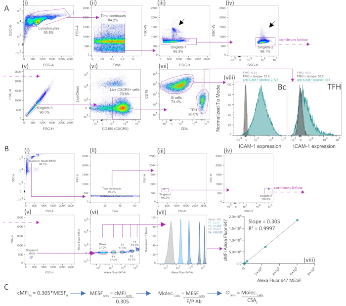
- Acquire the MESF standards, making sure the brightest peaks remain in the linear detection range for the quantification detector (channel), as shown in Figure 1B (vii).
- Acquire the samples manually or using HTS. If using the latter, resuspend BSLBs in 100 µL of PBS and acquire 80 µL using a flow rate between 2.5 and 3.0 µL/s, a mixing volume of 100 µL (or 50% of the total volume if the resuspension volume is less), a mixing speed of 150 µL/s, and five mixes to ensure BSLBs are monodispersed.
- Export the FCS files.
- Focus on single events for the analyses (Figure 2A), as doublets or triplets will introduce errors in the determination. Use nested identification of single events as indicated in protocol step 1.2.3.
- Measure the MFI of each MESF fraction (1-4) and subtract the MFI of blank beads to extract corrected MFIs (cMFI).
- Perform a linear regression analysis to extract the slope (b) of MESF over the cMFI calculated for MESF standards.
- Extract the MFI of each titration point and subtract the MFI of beads without protein to obtain cMFIs.
- Divide the cMFIs with the slope calculated to extract the MESF bound to BSLBs for each titration point.
- Divide MESF bound to BSLBs by the F/P value of the quantification antibody to extract the average number of molecules bound per BSLB.
- Using the diameter of the BSLBs (5.00 ± 0.05 µm), extract the bead surface area (SA = 4pr2) to calculate the final densities of protein (molec./µm2) per titration point (protein concentration).
- Perform a new regression analysis of protein concentration over protein density to calculate the slope (b) of the line of best fit.
NOTE: The concentration of 12.5 mol% of DGS-NTA(Ni) confers a maximum anchoring capacity of approximately 10,000 molec./µm2 10 without inducing the nonspecific activation of T cells or affecting the lateral mobility of the SLBs.
2. Performing synaptic transfer experiments between T cells and BSLBs
- Before running the synaptic transfer experiment
- Acquire non-fluorescent BSLBs, BSLBs with fluorescent lipids, unstained cells (or compensation beads; see the Table of Materials), and single-color-stained cells (or compensation beads) to identify the instrument's fluorescence spectrum interactions. Focus on those detectors with high spillover spreading to redesign the polychromatic panel, increase sensitivity, and reduce the measurement error on critical detectors.
- Titrate the detection antibodies to find the optimal concentration, enabling positive events detection without compromising the detection of negatives.
NOTE: Repeat this step whenever there is an antibody lot change as the F/P values and brightness vary from batch to batch.
- Optional: Optimize the PMT voltages by acquiring the sample at different voltage ranges (i.e., a voltage walks) to find the PMTs leading to optimal signal over noise (i.e., separation of negatives and positives while ensuring the signal of the brightest population remains in the linear range).
- Measurement of T-cell output transfer to BSLBs
- Prepare supplemented RPMI 1640 (herein R10 medium) containing 10% of heat-inactivated fetal bovine serum (FBS), 100 µM non-essential amino acids, 10 mM HEPES, 2 mM L-glutamine, 1 mM sodium pyruvate, 100 U/ml of penicillin, and 100 µg/mL of streptomycin. Use the R10 medium to culture and expand T cells.
- On day 1, isolate T cells from peripheral blood or leukoreduction system (LRS) chambers. Use immunodensity cell isolation and separation kits (see Table of Materials) for the enrichment of human CD4+ and CD8+ T cells.
- Seed the cells at a final concentration ranging between 1.5 and 2.0 × 10⁶ cells/mL, using 6-well plates with no more than 5 mL total per well.
- Activate T cells using a 1:1 ratio of human T cell activation (anti-CD3/anti-CD28) magnetic beads (see the Table of Materials) and add 100 IU of recombinant human IL-2 to support cell proliferation and survival.
- On day 3, remove the activating magnetic beads using magnetic columns (see the Table of Materials). Ensure that magnetic beads remain attached to the sides of the tube before recovering the cells for additional washing steps.
- Wash the magnetic beads once more with 5 mL of fresh R10 medium, mix well, and put them back in the magnet. Recover this volume and mix it with the cells recovered in step 1.2.5.
- Resuspend the cells to 1.5-2 × 10⁶ cells/mL in fresh R10 containing 100 U/mL of IL-2. Replenish the medium after 48 h, making sure that the last addition of IL-2 is 48 h before the day of experimentation (days 7 to 14 of culture).
- On the day of the synaptic transfer experiment, prepare the Synaptic Transfer Assay medium by supplementing Phenol Red-free RPMI 1640 medium with 10% FBS, 100 µM non-essential amino acids, 2 mM L-glutamine, 1 mM sodium pyruvate, 100 U/ml of penicillin, and 100 µg/mL of streptomycin. Do not include recombinant human IL-2.
- Count cells using either trypan blue staining or electric current exclusion and resuspend them to a final concentration of 2.5 × 10⁶ cells/mL in the medium. If excessive cell death is observed (>10%), remove dead/dying cells using a mixture of polysaccharide and sodium diatrizoate (see the Table of Materials) as follows:
- Layer 15 mL of cell culture on top of 13 mL of the polysaccharide-sodium diatrizoate solution.
- Centrifuge for 1,250 × g for 20 min at RT with minimum acceleration and deacceleration. Collect the cell layer (cloud) in the interface between the medium and the polysaccharide-sodium diatrizoate solution.
- Wash the cells at least twice with a prewarmed Synaptic Transfer Assay medium (prepared in step 1.2.7).
- Count and resuspend the cells to a final concentration of 2.5 × 10⁶/mL using a Synaptic Transfer Assay medium. Coculture 100 µL of this cell suspension with BSLBs (see step 1.2.15).
- Calculate the number of BSLBs needed for the experiment; consider all the different protein master mixes and antigen titrations to be tested (either biotinylated HLA/MHC-peptide monomers or monobiotinylated anti-CD3ε Fab).
- Assemble the BSLBs by following the same steps from protocol section 5 but this time, combine all the titrations of proteins and lipids required to reconstitute a complex APC membrane (Figure 3A). Keep the same vol: vol relationships between the initial silica beads volume and the volume of protein mix used during calibrations, as well as times and temperatures used in the loading of BSLBs. For example, if 0.5 µL of silica beads/well and 100 µL/well of protein mix were used for the initial calibration, maintain 5:1,000 vol:vol ratios to prepare the BSLBs to be cocultured with T cells.
- Once BSLBs have been loaded with the protein mix of interest, wash the BSLBs twice with HBS/HSA (BSA) to remove excess unbound proteins. Use sedimentation speeds of 300 × g for 2 min at RT in each washing step and discard the supernatants.
- Resuspend the 5 × 10⁵ BSLBs per well in 200 µL.
- Transfer 100 µL per well to a new U-bottom 96-well plate to make a duplicate, such that the final amount of BSLB per well is 2.5 × 10⁵.
- Spin down the BSLBs at 300 × g for 2 min and RT and discard the supernatant.
- Resuspend the BSLBs using 100 µL of T cell suspension; mix gently to prevent the formation of bubbles.
- Incubate the cocultures for 90 min at 37 °C.
NOTE: Alternatively, cells and beads can be resuspended in HBS/HSA (BSA) buffer instead of Phenol-Red free RPMI for the coculture. In this case, the incubation must be performed in a non-CO2 incubator as this gas will rapidly acidify the buffer in the absence of bicarbonate.
- Cool down the BSLB-T cell cocultures by first incubating the cells at RT for a minimum of 15 min. Protect them from light.
- Centrifuge the cells for 5 min at 500 × g and RT; discard the supernatant.
- Resuspend the cells in RT 2% BSA-PBS (Ca2+ and Mg2+-free) for blocking. Place the cells on ice for 45 min. Protect from light.
- While incubating the cells, prepare the antibody master mix using ice-cold 0.22 µm-filtered 2% BSA in PBS as a staining buffer, which will provide extra blocking.
NOTE: Some batches of antibodies conjugated to Brilliant Violet dyes tend to bind to BSLBs nonspecifically. Blocking with 5% BSA-PBS helps to reduce this noise. From now on, make sure to keep the cold chain unbroken.
- Spin down the cocultures at 500 × g for 5 min and 4 °C. Before discarding the supernatants, briefly make sure the pellet is present by inspecting the bottom of the 96-well plate using a backlight.
- Using a multichannel pipette, resuspend the cells in the staining master mix containing optimized antibody concentrations.
NOTE: Use pipette tips with no filter to prevent the generation of bubbles and errors in the distribution of staining volumes.
- Include isotype-labeled cells and BSLBs, fluorescent and non-fluorescent BSLBs, and cells and BSLBs stained alone. Respect the total number of events per well for all controls (i.e., only BSLBs containing 5 × 10⁵ BSLB/well, and only cell controls containing 5 × 10⁵ cells/well to avoid a relative increase of antibodies per stained event).
- Mix gently by pipetting up and down half of the volume and incubate for 30 min on ice. Protect from light.
- Wash the cells and BSLBs twice using ice-cold 2% BSA-PBS, pH 7.4, and sedimentation steps of 500 × g for 5 min at 4 °C. Resuspend the washed cocultures in 100 µL of PBS and acquire immediately.
- If fixation is needed, fix using 0.5% w/v of PFA in PBS for 10 min, wash once, and keep in PBS until acquisition. Protect from light.
- Before compensation, acquire MESF standards, ensuring both the dimmest and brightest populations fall in the linear range of measurement.
- Acquire compensation samples, calculate compensation, and apply the compensation matrix (link) to the experiment.
- Acquire and save a minimum of 2 × 10⁴ total MESF standards for each of the quantification channels.
- For acquisition using high-throughput samplers, set instrument acquisition to standard, set sample acquisition to 80 µL (or 80% of total volume), sample flow rate between 2.0 and 3.0 µL/s, sample mixing volume of 50 µL (or 50% of the total volume to avoid bubble formation during mixing), sample mixing of 150 µL/s, and mixing per well between 3 and 5.
- Acquire a minimum of 1 × 10⁴ single BSLBs per sample (refer to Figure 3B panels (i)-(vi) for the reference gating strategy).
- Wash the cytometer running for 5 min a cleaning solution followed by 5 min of ultrapure water before shutting down the instrument. If using the HTS, follow the options under the tab HTS and the Clean option.
- Export FCS files.
3. Measuring the synaptic transfer of particles to BSLBs
- Open the experiment FCS files. Select the population of cells and BSLBs based on their side and forward light scattering areas (SSC-A versus FSC-A), as shown in Figure 3B (i).
- Select the events within the continuous acquisition window (Figure 3B (ii)).
- Focus on the single events of both cells and BSLB; identify single cells first based on low W in the sequential gates FSC-W/FSC-H (singlets-1, Figure 3B panel (iii)) and SSC-W/SSC-H (singlets-2; Figure 3B panel (iv)). Define an additional singlets-3 gate by selecting events with proportional FSC-A and FSC-H (Figure 3B, panel (v)).
- Extract the MFI of MESF fractions blank and 1 to 4 and from single cells and MESF for each experiment sample.
- Generate corrected MFI (cMFI) values for MESF fractions 1 to 4 by subtracting the MFI of the blank bead population from each fraction.
- Generate cMFIs for single BSLBs and cells (refer to Figure 3C panel (i)).
- Use the signal from BSLB stained with isotype control antibodies to correct the MFI of BSLB stained with antibodies against the relevant T cell markers. Use cMFI to calculate the normalized synaptic transfer percent (NST%) by using the equation shown in Figure 3C panel (ii).
- If interested instead in the particles specifically transferred in response to TCR triggering, subtract the signal from null BSLBs from the MFI of agonistic BSLBs. Use this cMFI to calculate the TCR-driven NST% by using the equation shown in Figure 3C panel (ii).
- If interested in determining the total number of molecules transferred as particle cargo across the T cell-BSLB interface, acquire MESF benchmark beads using the same instrument settings and acquisition session for T cell-BSLB cocultures.
- Analyze MESF bead populations and extract their cMFIs. Calculate the slope of the line of best fit for the regression analysis of MESF over cMFI.
- Use the calculated slope to extract the number of MESF deposited on BSLBs. Use cMFIs calculated using either isotype controls or null BSLB as blanks to extract the number of MESF transferred specifically to stimulating BSLBs.
- Calculate the number of molecules of markers transferred to BSLBs by dividing the calculated (average) MESFs per BSLB by the F/P value of the quantification antibody.