1. Transfect mRNA to Cells
- Seed HeLa cells in a 24-well plate (to be approx. ~80-90% confluent the next day) and incubate overnight in an incubator at 37 °C with 5% CO2.
- Infect HeLa cells with vaccinia virus (VACV) at a Multiplicity of Infection (MOI) of 5 or keep uninfected HeLa cells for comparison.
NOTE: MOI is the number of infectious viral particles per cell.
- MOI of X= {[(Number of cells * X) / Virus Titer] * 1000} µl of virus per 1 mL medium.
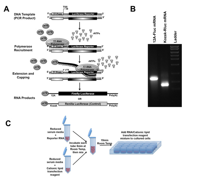
- After desired h post-infection (hpi) (in this experiment at 10-12 hpi), transfect mRNA (500 ng of total mRNA per well of 24-well plates) using a cationic lipid transfection reagent as shown in Figure 1C.
- For one well of a 24-well plate, mix 480 ng of 12A sequence bearing firefly luciferase (12A-Fluc) mRNA and 20 ng of Kozak sequence bearing Renilla luciferase (Kozak-Rluc) mRNA in one microcentrifuge tube. In another microcentrifuge tube add 1.1 µL of cationic lipid transfection reagent.
- Add 55 µL of reduced serum medium in both tubes. Mix and incubate at room temperature for 5 min.
- After 5 min of incubation, add 55 µL cationic lipid transfection reagent containing reduced serum medium in mRNA mRNA-containing tube.
- Mix gently but thoroughly, and incubate at room temperature for 15 min.
- During the incubation, remove the cell culture medium and add 400 µL of reduced serum medium per well of 24-well plates.
- After incubation, add 100 µL of the mixture dropwise and evenly to one well of 24-well plates.
2. Measure Luciferase Activities
- Five hours post-co-transfection of 12A-Fluc and Kozak-Rluc mRNA, measure luciferase activity using a luciferase assay system capable of performing two reporter assays (e.g., Dual-Luciferase Reporter assay kit).
- Remove the reduced serum medium and lyse the cells by adding 150 µL 1x lysis buffer, a component of the luciferase assay kit.
- After 10 min incubation at room temperature, collect the lysate by scrapping the cells and transfer them to a microcentrifuge tube.
- Centrifuge the lysate at 12,000 x g for 10 min at 4 °C to pellet cell debris.
- Add 30 µL of supernatant in an opaque-walled 96-well white assay plate with a solid bottom.
- Measure the dual luminescence using the luciferase assay kit and a multimode plate reader luminometer.
- Perform the measurement using the kinetics function (on a per-well basis) using the settings described in Table 1.
NOTE: The reading can also be taken using a manual luminometer. Add an equal volume of lysate and substrate for Fluc in a cuvette. Wait for 2 s and measure for 10 s using a luminometer. Following the Fluc measurement, quickly take out the cuvette from the luminometer and add an equal volume of the substrate for Rluc manually. Again, wait for 2 s and measure for 10 seconds using a luminometer.
- Export the luminescence reading data into a desirable file format.
- Determine the relative translation rate from 12A-Fluc mRNA in uninfected and VACV-infected HeLa cells by dividing the Fluc value by the internal control Rluc value.
NOTE: Figure 2 shows the step-by-step analysis of raw data to get relative Fluc activity.
Table 1: Luciferase Measurement Settings – the steps for luciferase measurement with the recommended volume or time.
| Steps | Volume/Time |
| Inject Luciferase Assay Substrate (Fluc): | 30 µL |
| Wait / Incubation time: | 2 s |
| Luminescence Measurement (Fluc): | 10 s |
| Stop & Glo Substrate (Rluc): | 30 µL |
| Wait / Incubation time: | 2 s |
| Luminescence Measurement (Rluc): | 10 s |