A subscription to JoVE is required to view this content. Sign in or start your free trial.

Method Article

A Technique to Assess the Inhibitory Effect of Toxin Exposure on Miniature Excitatory Postsynaptic Currents (mEPSCs)

904 views

July 8th, 2025

In This Article

Abstract

Source: Hubbard, K. et. al., Functional Evaluation of Biological Neurotoxins in Networked Cultures of Stem Cell-derived Central Nervous System Neurons. J. Vis. Exp. (2015)

The video demonstrates the electrophysiological analysis of embryonic stem cell-derived neurons after exposure to neurotoxins. Treating these neurons with botulinum neurotoxins reduces the frequency of spontaneous miniature excitatory post-synaptic currents, confirming inhibition of synaptic transmission.

Protocol

1. Adaptation of embryonic stem cells (ESCs) to Feeder Cell-free Suspension Culture

  1. Thaw ESCs at 37 °C until a sliver of ice remains.
  2. Using a P1000 pipet, gently transfer 1 - 2.5 x 10⁶ dissociated ESCs to a non-tissue culture-treated dish containing 10 ml of pre-warmed ESC medium. Incubate in a humidified tissue culture incubator for 20 hr at 37 °C and 5% carbon dioxide (CO2).
  3. After 20 hr, wash cells by gently transferring the suspension culture to a 15 ml conical tube using a 10 ml serological pipet. Pellet ESCs for 3 min at 200 x g. During spin, add 10 ml of fresh ESC medium to low-adhesion dish.
  4. Aspirate supernatant, taking care to avoid dislodging the cell pellet, and add 1 ml fresh ESC medium to the cell pellet. Transfer cells to the bacterial dish using a P1000 pipet and return to the incubator.
  5. Observe cells daily until aggregates become visible to the naked eye (typically 4 - 8 days (d); aggregates will be 0.2 - 0.5 mm). If aggregates are not apparent after 4 d, repeat step 1.3. Once aggregates are visible, dissociate and maintain ESCs as described in Section 2.

2. ESC Passaging and Maintenance

  1. Transfer ESC aggregates to a 15 ml conical tube using a sterile 10 ml pipet and allow aggregates to settle to a compact pellet (typically 3 - 5 min). Aspirate the supernatant, being careful to avoid disrupting the cell pellet, and wash aggregates with 5 ml phosphate-buffered saline (PBS). Although gravity settling is recommended, alternatively, pellet cells at 100 x g for 2.5 min instead to save time.
  2. Aspirate PBS and add 500 µl trypsin. Incubate aggregates in trypsin for 3 min in a water bath at 37 °C. Add 500 µl ESC medium to dilute the trypsin and gently triturate 10 times with a P1000 pipet to break up aggregates. Count cells using a hemocytometer.
  3. Pellet dissociated cells for 3 min at 200 x g and resuspend cells to a final concentration of 1.0 x 10⁷ cells/ml in ESC medium. Transfer 150 µl (1.5 x 10⁶ cells) to 10 ml of fresh ESC medium in a 10 cm bacterial dish and return to tissue culture incubator for 48 hr. Excess ESCs can be differentiated, cryopreserved at 1 - 5 x 10⁶ cells/ml in 90% ESC medium supplemented with 10% dimethyl sulfoxide (DMSO), or discarded.
  4. Passage aggregates every 48 hr. Aggregates ready for passage will be clearly visible, with diameters exceeding 0.5 mm.
    NOTE: Thawing and culture of cryopreserved, suspension-adapted cells are accomplished per steps 1.2 - 1.4. Aggregates will be ready for passaging within 48 - 72 hr.

3. Neuronal Differentiation

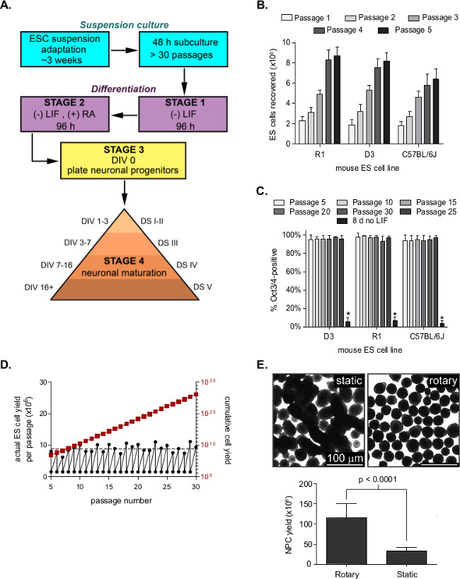
NOTE: Conduct steps 3.1 - 3.5 before noon and step 3.7 after noon. An overview of the differentiation procedures is presented in Figure 1A.

  1. Dissociate ESCs as in steps 2.1 - 2.5. Transfer 350 µl (3.5 x 10⁶) of dissociated ESCs to a 10 cm low-attachment dish containing 25 ml differentiation medium. Place on an orbital shaker set to 30 - 45 rpm inside a tissue culture incubator at 37 °C with 5% CO2. This is the first day of differentiation, termed day in vitro (DIV) -8.
    NOTE: Using an ultra-low attachment dish increases the cost of the method but produces slightly larger yields than bacterial Petri dishes since aggregates can occasionally adhere to Petri dishes. Medium volumes and cell numbers can be scaled accordingly if different dish sizes are preferred.
  2. After 48 hr (DIV -6), use a 25 ml pipet to transfer the differentiating cell aggregates to a 50 ml conical tube. Immediately add a fresh 25 ml of differentiation medium to the Petri dish.
  3. Allow aggregates to settle over 2 - 5 min, producing a visible pellet that is 1 - 2 mm deep. Ignore single cells or small aggregates remaining in suspension. Carefully aspirate the medium and transfer the cell pellet back to the petri dish using a P1000. Place on rotary shaker in a tissue culture incubator.
  4. At DIV -4, repeat step 3.2. The pellet will be 2 - 4 mm deep. Replace 30 ml differentiation medium supplemented with 6 µM all-trans retinoic acid (RA) to the petri dish. Return to the rotary shaker in the tissue culture incubator for an additional 48 hr.
  5. At DIV -2, repeat step 3.3. The pellet will be 4 - 8 mm deep at this point.
  6. At DIV -1, prepare plating surfaces as in Section 4.
  7. At DIV 0, thaw 5 ml of pre-aliquoted and frozen neural progenitor cells (NPC) trypsinization medium at 37 °C for 5 - 10 min and place in a tissue culture hood. Using a 25 ml pipet, transfer differentiating aggregates to a 50 ml conical tube. Allow aggregates to settle and carefully aspirate the medium. Wash the pellet twice with 10 ml PBS, allowing the aggregates to settle between washes.
  8. After the second PBS wash, add 5 ml of NPC trypsinization medium to the pellet and incubate at 37 °C for 5 min. Gently flick the tube after 2.5 min.
  9. Add 5 ml of 0.1% soybean trypsin inhibitor (STI) to inactivate trypsin and mix by inverting. Gently triturate 10 - 15 times with a 10 ml serological pipet until a relatively homogenous cell suspension is produced.
  10. Slowly transfer the cell suspension to a 40 µm or 70 µm cell strainer placed on the top of a 50 ml conical tube. Once all the suspension has been filtered, add 1 ml of N2 medium to wash the remaining cells through the filter and pellet the dissociated cell suspension for 6 min at 200 x g.
  11. Aspirate medium without disturbing the pellet. Wash cells twice with 10 ml N2 medium, pellet cells for 5 min at 200 x g, and triturate between washes with a P1000. Before the second wash, count cells using a hemocytometer.
  12. Resuspend cells in N2 medium at 1 x 10⁷ cells per ml and plate embryonic stem cell-derived neurons (ESNs) at a cell density of 150,000 - 200,000 cells/cm2.
  13. Transfer newly plated ESNs to a humidified tissue culture incubator at 37 °C and 5% CO2 and maintain as in Section 5.

4. Preparing Culture Surfaces for Plating Neural Precursors at DIV 0

  1. Prepare tissue culture-treated dishes at least 1 day before plating. Add sufficient polyethyleneimine (PEI; 25 µg/ml in sterile H2O) or poly-D-lysine (PDL; 100 µg/ml in sterile H2O) to cover tissue-culture-treated plastic dishes and incubate O/N at 37 °C.
  2. The morning of plating, wash dishes twice with double-distilled H2O and once with PBS. After the final wash, add sufficient N2 medium to cover the dish (e.g., 1 ml per well of a 12-well dish or 4 ml per 6 cm dish).
  3. Prepare 18 mm glass coverslips at least one day before neuron plating. Clean coverslips by plasma-cleaning for 4 min.
  4. Immediately transfer cleaned coverslips to an ethanol-washed parafilm in the bottom of a large sterile dish and add 400 µl of PEI or PDL solution, prepared as in step 4.1. Incubate overnight (O/N) at 37 °C in a tissue culture incubator.
  5. In the morning, wash coverslips three times with water and add 5 µg/ml laminin in PBS for 1 - 3 hr at 37 °C. Before dissociating neurons, aspirate the laminin and immediately transfer the coverslip to a well of a 12-well dish containing 1 ml of NPC medium, being sure to keep the treated side facing up.
  6. Store dishes and coverslips at 37 °C until NPCs are ready to plate.

5. Maintenance of Neurons

  1. At DIV 1, aspirate medium and replace with N2 culture medium.
  2. At DIV 2 and 4, aspirate medium and replace with B27 culture medium.
  3. At DIV 8, aspirate medium and replace with B27 culture medium containing mitotic inhibitors to eliminate contaminating non-neuronal cells.
  4. At DIV 12, replace with B27 culture medium.
  5. Do not remove DIV 12+ ESNs from 5% CO2 until ready.

6. Measured Inhibition of Synaptic Transmission (MIST) Assay for Quantifying Miniature Excitatory Post-synaptic Currents

CAUTION: Clostridial neurotoxins are the most poisonous substances known, with estimated human LD50 values as low as 0.1 - 1 ng/kg. Obtain necessary approvals before using these toxins and use appropriate precautions.

  1. Carefully dilute BoNT/A to 100x, the desired final concentration in ESN culture medium, and warm to 37 ºC. Add the appropriate volume of toxin to DIV 21+ ESN cultures, swirl the culture dish, and return to the incubator.
  2. If cells will be analyzed more than 4 hours after intoxication, add a toxin to the dish and swirl without removing from 5% CO2, such as directly in the incubator or in a constant CO2 chamber.
  3. At the appropriate time point, aspirate the ESN culture medium and wash twice with extracellular recording buffer (ERB). Add 4 ml of ERB supplemented with 5.0 µM tetrodotoxin and 10 µM bicuculline, blocking action potentials and antagonizing GABAA receptor activity, respectively.
  4. Transfer the dish to the electrophysiology rig. Neither perfusion nor temperature control is required for the MIST assay.
  5. Pull borosilicate glass using a micropipette puller to produce a recording pipette with 5-10 mΩ of resistance and fill with intracellular recording buffer. Gently dip filled recording pipette in siliconizing reagent before recording.
  6. Using an air-filled syringe, provide positive pressure as the recording pipette is lowered into the ERB. After gently landing the recording pipette on the neuron's soma to be recorded, remove positive pressure and form a gigaseal. Decrease the holding voltage to -70 mV. Carefully apply negative pressure to break into whole-cell configuration.
  7. Switch to the current clamp to monitor and record resting membrane potential. Without adjustment for liquid junction potentials, the resting membrane potential will be between -67 and -82 mV.
  8. Perform a continuous -70 mV voltage-clamp recording for 4 - 5 min to detect miniature excitatory post-synaptic currents (mEPSCs).
  9. Analyze 4 min of recorded data for mEPSC detection using spike detection software with the following settings: Threshold, 5; Period to search a local maximum, 10,000 µsec; Time before a peak for baseline, 5,000 µsec; Period to search a decay time, 20,000 µsec; Fraction of peak to find a decay time, 0.37; Period to average a baseline, 1,000 µsec; Area threshold, 20; Number of points to average peak, 1; Direction of peak, negative.
  10. Collect and save information on detected events. Divide the number of detected events in 4 min by 240 to determine the mEPSC frequency in Hz.
  11. Collect mEPSC frequencies for 8 - 12 controls and 8 - 12 BoNT-treated samples for each exposure condition. Analyze frequency against age- and lot-matched controls. Determine the statistical significance of % inhibition of synaptic activity using one-way ANOVA and Dunnett's post-hoc test.

Access restricted. Please log in or start a trial to view this content.

Results

Embryonic stem cell differentiation process, charts, graphs, and microscopy for cell line analysis.
Figure 1. Suspension-adapted ESNs remain mitotically stable and express markers of pluripotency. (A) Schematic of ESC maintenance and differentiation. The presence or absence of retinoic acid (RA) or leukemia inhibitory factor (LIF) is m...

Access restricted. Please log in or start a trial to view this content.

Disclosures

No conflicts of interest declared.

Materials

List of materials used in this article
NameCompanyCatalog NumberComments
Table 1. ESC and ESN
Knockout DMEMLife Technologies10829-018Media: ESC culture medium
Formulation and notes: 500 mL
100x MEM NEAALife Technologies11140-050Media: store at 4 °C for up to 1 month
Formulation and notes: 6 mL
200 mM L-Alanyl-L-GlutamineATCC30-21156 mL
ES qualified FBSApplied Stem CellASM-500790 mL
100x antibioticsSigma-AldrichA59553 mL
55 mM 2-mercaptoethanolLife Technologies21985-0231.1 mL
107 Units/mL LIFMilliporeESG110760 μL
Knockout DMEMLife Technologies10829-018Media: ESC differentiation medium
Formulation and notes: 436.6 mL
100x MEM NEAALife Technologies11140-050Media: store at 4 °C for up to 1 month
Formulation and notes: 5 mL
200 mM L-Alanyl-L-GlutamineATCC30-21155 mL
ESC-qualified serumApplied Stem CellASM-500750 mL
100x antibioticsSigma-AldrichA59552.5 mL
55 mM 2-mercaptoethanolLife Technologies21985-0230.9 mL
Dulbecco's PBSSigma-AldrichD8537Media: NPC trypsinization medium
Formulation and notes: 100 mL
0.5 M EDTA (18.3%)Sigma-Aldrich3690Media: freeze in 5 mL aliquote
Formulation and notes: 0.266 mL
TrypsinSigma-AldrichT880250 mg
Polyethyleneimine (PEI)Sigma-AldrichP3143Media: Surface coating solutions
Formulation and notes: 2.5 µg/mL in H20
Poly-D-lysine (PDL)Sigma-AldrichP7280Media: use within 1 wk
Formulation and notes: 100 µg/mL in H20
LamininSigma-AldrichL20205 µg/mL in H20
DMEM/F12 + GlutaMAXLife Technologies10565-018Media: NPC culture medium
Formulation and notes: 492.5 mL
100x N2 vitaminsLife Technologies17502-048Media: store at 4 °C for up to 1 month
Formulation and notes: 5 mL
100x antibioticsSigma-AldrichA59552.5 mL
Neurobasal ALife Technologies10888-022Media: ESN culture medium
Formulation and notes: 482.5 mL
50x B27 vitaminsLife Technologies17504-044Media: store at 4 °C for up 1 month
Formulation and notes: 10 mL
200 mM L-Alanyl-L-GlutamineATCC30-21155 mL
100x antibioticsSigma-AldrichA59552.5 mL
5-fluoro-2'-deoxyuridine (5FDU)Sigma-AldrichF0503Media: 2000x Mitotic inhibitors aliquot and store at -20 °C
Formulation and notes: dd 150 mg 5FDU and 350 mg uridine to 10 mL Neurobasal A. Sterile filter, aliquot and freeze. Use 2000x in ESN culture medium.
UridineSigma-AldrichU3003
TrypLE Express TrypsinLife Technologies12605-010Media: Miscellaneous
Formulation and notes: store at RT
DMSOSigma-AldrichD8418store at RT
Soybean trypsin inhibitor (STI)Sigma-AldrichT6414store at -20 °C
EthanolSigma-AldrichE7023store at RT
Ascorbic acidSigma-AldrichA4403Prepare 100 mM stock by dissolving 100 mg ascorbic acid in 5.7 mL of 50:50 DMSO/ethanol mix.
Retinoic acid (RA)Sigma-AldrichR2625Resuspend 15 mg RA in 9 mL 50:50 DMSO/ethanol. Supplement with 1 mL of 100 mM ascorbic acid stock. Aliquot and stored at -80 °C. Stable for 6 mos.
Table 2. MIST
NaClSigma-Aldrich71386Media: Extracellular Recording Buffer
Final Concentration (mM): 58.44
MW: 140
KClSigma-Aldrich60135Media: (ERB; pH = 7.3, 315 mO)
Final Concentration (mM): 74.56
MW: 3.5
NaH2PO4Sigma-AldrichS3139Media: Stable at 4 °C for at least 6 mo.
Final Concentration (mM): 119.98
MW: 1.25
CaCl2Sigma-Aldrich21115Final Concentration (mM): 110.98
MW: 2
MgCl2Sigma-Aldrich63069Final Concentration (mM): 95.21
MW: 1
D-glucoseSigma-AldrichG8644Final Concentration (mM): 180.16
MW: 10
HEPESSigma-AldrichH0887Final Concentration (mM): 238.3
MW: 10
K-gluconateSigma-AldrichP1847Media: Intracellular Recording Buffer
Final Concentration (mM): 234.5
MW: 140
NaClSigma-Aldrich71386Media: (IRB; pH = 7.3, 320 mO)
Final Concentration (mM): 58.44
MW: 5
Mg-ATPSigma-AldrichA9187Media: Stable at 4 °C for at least 6 mo.
Final Concentration (mM): 507.18
MW: 2
Li-GTPSigma-AldrichG5884Final Concentration (mM): 523.18
MW: 0.5
CaCl2Sigma-Aldrich21115Media: Stable at 4 °C for at least 6 mo.
Final Concentration (mM): 110.98
MW: 0.1
MgCl2Sigma-Aldrich63069Media: Stable at 4 °C for at least 6 mo.
Final Concentration (mM): 95.21
MW: 1
EGTASigma-AldrichE3889380.35
Media: Stable at 4 °C for at least 6 mo.
Final Concentration (mM): 380.35
MW: 1
HEPESSigma-AldrichH0887Media: Stable at 4 °C for at least 6 mo.
Final Concentration (mM): 238.3
MW: 10
BicucullineTocris131Media: Miscellaneous
Final Concentration (mM): 0.01
MW: 417.85
CNQXSigma-AldrichC239Final Concentration (mM): 0.01
MW: 276.12
TetrodotoxinSigma-AldrichA8001Final Concentration (mM): 0.005
MW: 645.74
BoNT/A-/GMetabiologicsN/AMedia:
Final Concentration (mM): TBD
MW: 150,000
Tetanus toxinSigma-AldrichT3194Final Concentration (mM): TBD
MW: 150,000
SigmacoteSigma-AldrichSL-2Final Concentration (mM): N/A
MW: N/A
Table 3. Equipment and Software
MiniAnalysis (version 6.0.7)Synaptosoft, Inc.N/AEvent detection software
Igor Pro (version 6.22A)WaveMetricsN/ASoftware for visualization of recordings
PatchmasterHekaN/AData acquisition software
Olympus IX51 inverted microscopeOlympusN/A
MicromanipulatorSutter InstrumentMPC-365
Patch-clamp amplifierHekaECB10USB
Micropipette pullerSutter InstrumentP-1000
Borosilicate capillary tubesSutter InstrumentB150-86-10Corning 7740
18 mm glass coverslipsFisher12-545-84
Plasma cleanerHarrick PlasmaPDC-32G
Cell strainer (40 µm)Fisher08-771-1
Stovall Belly Dancer ShakerFisher15-453-211
Low adhesion dishesFisher05-539-101Corning 3262
Bacterial dishesVWR25384-302100 x 15 mm

Tags

Botulinum Neurotoxin ExposureElectrophysiological AnalysisStem Cell derived NeuronsNeurotransmitter Release InhibitionGigaseal FormationVoltage clamp RecordingTetrodotoxin ApplicationBicuculline AntagonismmEPSC Frequency Measurement