Method Article

Measuring the Activation of Fc-Mediated Effector Functions by HA Antibodies

July 8th, 2025

In This Article

Abstract

Loading...
$$\rightleftharpoonup{xx}$$ $$\longleftharp{xx}$$, $$\longrightharp{xx}$$,

Source: Bailey, M. J., et al. A Method to Assess Fc-mediated Effector Functions Induced by Influenza Hemagglutinin Specific Antibodies. J. Vis. Exp. (2018).

This video demonstrates an assay measuring the antibody-mediated activation of T cell effector functions. Adding an influenza virus hemagglutinin (HA)-specific monoclonal antibody to transfected mammalian cells expressing HA results in the formation of immune complexes with HA. Upon introducing engineered T cells expressing Fc receptors and a luciferase reporter, the T cells are activated by the antibody-HA complexes, and this activation is detected by adding a luciferase substrate and measuring the produced bioluminescence.
Protocol

Loading...
$$\rightleftharpoonup{xx}$$ $$\longleftharp{xx}$$, $$\longrightharp{xx}$$,

1. Expression of Influenza Virus Hemagglutinin via Transfection

  1. Plate Human Embryonic Kidney (HEK 293T) cells at a density of 2 x 10⁴ cells/well in a white tissue culture treated 96-well plate and let it sit for 4 h in a 37 °C incubator (with 5% CO2).
  2. Transfect cells with 100 ng of DNA coding for viral hemagglutinin and 0.2 μL of transfection reagent per well in a total volume of 50 μL.
    NOTE: Both plasmid and transfectant reagent are diluted in 1x Opti-Minimum Essential Media (MEM) Reduced Serum Medium.
  3. After incubating plates in a 37 °C incubator with 5% CO2 for 18 h, remove transfection media.

2. Assessing the FcRg/NFAT-mediated Activation of Luciferase Activity by Monoclonal Antibodies or Sera

  1. First, replace the growth media of transfected cells with 25 µL of assay media (1x RPMI 1640 supplemented with 4% (vol/vol) low IgG fetal bovine sera).
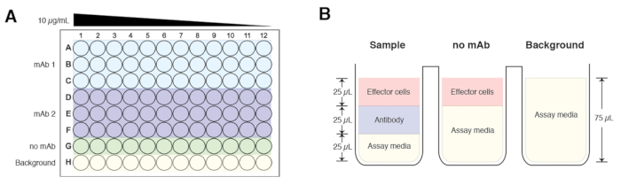
  2. In a separate 96-well plate, perform four-fold dilutions of the antibodies of interest starting at 30 μg/mL using the assay media. Perform dilutions in triplicates, and an example plate layout is illustrated in Figure 1A. Ensure that a control that excludes antibodies (no antibody control) and a background control (assay buffer alone) is included.
    NOTE: For both the ‘no antibody' and background controls, add 25 µL of assay buffer instead of the antibody.
  3. Transfer 25 μL of the serially diluted antibodies from Step 2.2 to corresponding wells on the plate of transfected cells.
  4. Incubate plate in a 37 °C incubator (with 5% CO2) for 15 min.
  5. Add 25 μL of the appropriate modified effector Jurkat cell expressing murine FcgRIV or human FcgRIIIa (75,000 cells/well). The total volume for each well should be 75 µL, and the starting final concentration of the antibody is at 10 µg/mL (Figure 1B).
    NOTE: For the background control, add 25 µL of assay buffer instead of effector cells. To examine antibody-dependent cell-mediated phagocytosis, a modified Jurkat cell expressing the human FcgRIIa can be used.
  6. Incubate the plate in a 37 °C incubator with 5% CO2 for 6 h.
  7. Thaw and warm luciferase substrate buffer in 37 °C water for about 1 h before use.
    NOTE: To make the luciferase substrate buffer, reconstitute the lyophilized luciferase assay substrate in the provided assay buffer. The reconstituted substrate buffer can be stored at -30 °C to -10 °C for up to six weeks.
  8. Add 75 μL/well of luciferase substrate buffer to all wells except for the background control.
  9. Read on a luminometer.
  10. Calculate fold induction with the following equation: Fold induction = (Induced value-Background value) / (No mAb value-Background value).

Access restricted. Please log in or start a trial to view this content.

Results

Loading...
$$\rightleftharpoonup{xx}$$ $$\longleftharp{xx}$$, $$\longrightharp{xx}$$,

Plate assay diagram and setup for antibody titration; shows sample, no mAb, and background controls.
Figure 1: A schematic of a sample plate layout and composition of the experimental groups. (A) The starting concentration for each antibody sample is at 10 µg/mL and serially diluted across the plate (column 1 to column 12) 4-fold....

Access restricted. Please log in or start a trial to view this content.

Disclosures

Loading...
$$\rightleftharpoonup{xx}$$ $$\longleftharp{xx}$$, $$\longrightharp{xx}$$,
No conflicts of interest declared.

Materials

List of materials used in this article
NameCompanyCatalog NumberComments
HEK 293T cellsATCCCRL-3216Human embryonic kidney cells
Lipofectamine 2000Life Technologies11668019Transfection reagent
1X Opti-MEM Reduced Serum MediumThermoFisher Scientific51985-034
White tissue culture treated 96-well plateCorning, Inc.3917Assay plates
10X MEMGibco11430-030
Jurkat cell expressing murine FcgRIVPromegaM1201Kit provides includes cells, Bio-Glo Luciferase assay system, 1X RPMI and low IgG serum
Jurkat cell expressing human FcgRIIIaPromegaG7010Kit provides includes cells, Bio-Glo Luciferase assay system, 1X RPMI and low IgG serum
Jurkat cell expressing human FcgRIIaPromegaG9901Kit provides includes cells, Bio-Glo Luciferase assay system, 1X RPMI and low IgG serum
LuminometerBioTekSynergy H1 Multi-Mode readerLuminescence plate reader
Bio-Glo Luciferase Assay SystemPromegaG7940Contains luciferase assay buffer and luciferase assay substrate

Reprints and Permissions

Request permission to reuse the text or figures of this JoVE article

Request Permission

Tags

HA Antibody AssayImmune Complex FormationT Cell ActivationLuciferase Reporter SystemNFAT Signaling PathwayAntibody Dependent Cell Mediated CytotoxicityBioluminescence DetectionHemagglutinin Stalk TargetingEngineered Jurkat Cells

Related Articles