Method Article

An Immunofluorescence-Based Method for Visualizing Tuft Cells in Jejunum Cryosections

July 8th, 2025

In This Article

Abstract

Loading...
$$\rightleftharpoonup{xx}$$ $$\longleftharp{xx}$$, $$\longrightharp{xx}$$,

Source: Mizutani, Y. et al., Use of Anti-phospho-girdin Antibodies to Visualize Intestinal Tuft Cells in Free-Floating Mouse Jejunum Cryosections. J. Vis. Exp. (2018)

This video illustrates an immunofluorescence-based method for visualizing intestinal tuft cells in free-floating mouse jejunum cryosections. Within the intestinal villi, the co-localization of green fluorescence at the luminal tip with an extended red rootlet mass indicates the presence of tuft cells.
Protocol

Loading...
$$\rightleftharpoonup{xx}$$ $$\longleftharp{xx}$$, $$\longrightharp{xx}$$,

All procedures involving animal models have been reviewed by the local institutional animal care committee and the JoVE veterinary review board.

1. Preparations

  1. Prepare 10 L of phosphate-buffered saline (PBS): 32.27 g of sodium hydrogen phosphate (Na2HPO4·12H2O), 4.5 g of Sodium dihydrogen phosphate (NaH2PO4·2H2O), 80.0 g of sodium chloride (NaCl) added to ultrapure water to a final volume of 10 L. Store solution at room temperature (RT).
  2. Prepare 200 mL of PBS-T: 200 mL of PBS from step 1.1 with 100 µL of polyoxyethylene (10) octylphenyl ether to a final concentration of 0.05% (vol: vol). Store at RT.
  3. While wearing gloves and eye protection, prepare 50 mL of 15% sucrose in 10% buffered neutral formalin solution: 7.5 g of sucrose added to 10% buffered neutral formalin solution (Table of Materials) to a final volume of 50 mL. Store at RT.
    CAUTION: Inhalation and/or skin/eye contact with formaldehyde in 10% buffered neutral formalin solution can be hazardous. Handle with caution.
  4. Prepare 1.5 mL of 200 units/mL phalloidin-fluorescent dye conjugate stock solution: phalloidin-fluorescent dye conjugate (300 units in 1 vial, lyophilized solids, excitation, and emission wavelengths: 581 nm and 609 nm, respectively) suspended in 1.5 mL of methanol. Store the solution in the dark at -20 °C.
  5. Prepare 5 mg/mL 4',6-diamidino-2-phenylindole, dihydrochloride (DAPI) stock solution: 10 mg of DAPI in 2 mL of N, N-dimethylformamide (DMF). Store the solution in the dark at -20 °C.
  6. Prepare 5% bovine serum albumin (BSA) in PBS: 0.5 g of BSA, 10 mL of PBS. Store at 4 °C.
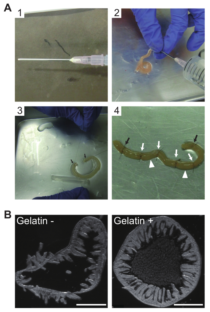
  7. Remove the beveled tip of an 18-gauge straight needle using a nipper, and pinch the nipped end with a pincher in a lengthwise direction to allow liquid to flow through the needle lumen (Figure 1A1).
    NOTE: The protocol can be paused here.

2. Animal Dissection and Isolation of Jejunum

  1. Perfusion fixation of a mouse
    1. Wear gloves and work in a well-ventilated area.
    2. Prepare a 100 mL glass beaker, 10% buffered neutral formalin solution, surgical tools (scissors, forceps), a metallic tray, 6-0 nylon cut sutures, an 18-gauge straight needle prepared as in 1.7, a 22-gauge winged needle, a 20-mL syringe.
    3. Load 20 mL of 10% buffered neutral formalin solution from the glass beaker into the 20-mL syringe and attach the 22-gauge winged needle to the outlet.
    4. Prepare liquid gelatin by adding 1 g of gelatin powder to 20 mL of PBS (final 5% gelatin) in a 50-mL centrifuge tube. After soaking at RT for 15 min, incubate at 50 °C in a water bath for 15 min without shaking.
    5. Briefly vortex and incubate at 50 °C for another 15 min to completely dissolve the gelatin. Leave the tube at RT until use.
    6. Euthanize an adult mouse by cervical dislocation.
    7. Place the mouse from step 2.1.6 on a metallic tray and make a small incision on the skin through the ensiform cartilage using surgical tools.
    8. Expand the incision with both hands to expose the thoracic and abdominal regions.
    9. Open the peritoneum to expose the diaphragm, and then open the diaphragm on both sides.
    10. Cut the thoracic cage along the anterior axillary lines on both sides, then remove the thoracic wall by cutting in a transverse direction at the position of the thymus to expose the heart.
    11. Make a small incision on the auricle of the right atrium to drain the blood and insert a 22-gauge winged needle into the apex of the left ventricle. Push the plunger to perfuse the tissues with a 10% buffered neutral formalin solution.
    12. After exposing organs in the pelvis, cut off the end of the rectum from the anus, and separate the intestines from the body by cutting the mesentery.
    13. To isolate the jejunum, cut the intestines at 4 cm from the anal side of the gastric antrum. Discard the anal half of the remaining small intestine.
    14. Clip the jejunum in half to facilitate the flushing procedure. Load 20 mL of 10% buffered neutral formalin solution into the 20-mL syringe, and attach the 18-gauge straight needle prepared in step 1.7 to the outlet.
    15. Inject 10% buffered neutral formalin solution from one end of the clipped jejunum to flush out the intestinal contents and to fix the gut lumen surface (Figure 1A2).
    16. Wash the gut lumen by injecting PBS as described in step 2.1.15.
    17. Flush liquid gelatin into the gut lumen as described in step 2.1.15 to replace PBS with liquid gelatin.
    18. Close one end of the clipped jejunum by suture ligation using a 6-0 nylon cut suture, fill the jejunum with liquid gelatin, and close the opposite end by suture ligation (Figure 1A3).
    19. Add four suture knots to fit a sausage-shaped jejunum section to the depressed portion of a cryomold (Figure 1A4).
      NOTE: For cryomolds with a 20 mm x 25 mm x 5 mm depressed section, a ~20 mm sausage-like jejunum section is preferable.
    20. Soak the tissues in 50 mL of 15% sucrose in 10% buffered neutral formalin solution overnight at 4 °C.
      NOTE: These steps ensure cryoprotection by sucrose and protein fixation by formalin of gelatin and tissue. Formalin-gelatinized gelatin does not melt at RT, whereas non-fixed gelatinized gelatin at 4 °C melts at RT.

3. Snap Freezing of Gelatin-filled Jejunum Tissues

  1. By cutting the jejunum at the suture knots, three sausage-like jejunum pieces with both ends ligated are obtained (Figure 1A4). Align the sausage-like pieces in a cryomold for the addition of embedding compound for the preparation of frozen tissue specimens.
  2. Snap freeze the cryomold in isopentane cooled with liquid nitrogen. Store cryomolds at -80 °C
    NOTE: The protocol can be paused here.

4. Immunofluorescence of Tuft Cells Using Free-floating Cryosections

  1. Cryosectioning
    1. Set both the cryostat chamber temperature (CT) and object temperature (OT) to -22 °C. Place the frozen tissue block in a cryomold in the cryostat chamber for at least 15 min.
    2. Add 3 mL of PBS to a 35-mm culture dish.
    3. Remove the frozen tissue block from the cryomold and cut the block in half with a razor blade to expose the transverse section of the jejunum. Mount one half of the block on a chuck (a cryostat adaptor) to section the cutting plane with the razor blade.
    4. Section the gelatin-filled jejunum into 30 µm-thick sections. Use frozen forceps to gently transfer the sections into the 35-mm culture dish prepared in step 4.1.2 (Figure 2B, right).
  2. Application of primary antibodies
    1. Wash the free-floating sections in the 35-mm culture dish 3 times for 5 min each with 3 mL PBS-T with mild shaking on a reciprocal shaker (approximately 36 times/min).
    2. Prepare antigen retrieval solution: 0.3 mL of antigen retrieval solution concentrate (Table of Materials) in 2.7 mL of ultrapure water. Add 3 mL of antigen retrieval solution to the 35-mm culture dish containing free-floating sections.
    3. Close the lid, seal the gap between the dish and the lid with a strip of vinyl tape, and incubate at 50 °C in a hybridization incubator for 3 h without shaking.
    4. Remove the dish from the incubator and cool at RT for 20 min. After removing the vinyl tape, wash the sections 3 times for 5 min each with 3 mL of PBS-T and mild shaking.
    5. Aspirate the PBS-T and add 5 drops of blocking solution (Table of Materials) onto the sections. Incubate at RT for 5 min with mild shaking.
    6. Prepare the primary antibody solution: 495 µL of 5% BSA in PBS with 5 µL of phospho-Y1798 girdin (pY1798) antibodies (Table of Materials). Add 500 µL of primary antibody solution onto the sections (the blocking solution does not need to be removed).
    7. Place the dish in a humidified incubation chamber and incubate overnight at 4 °C with mild shaking.
      NOTE: The overnight incubation can be extended to up to three nights.
  3. Application of secondary antibodies
    1. Wash the sections 3 times for 5 min each in 3 mL of PBS-T with mild shaking.
    2. Prepare diluted DAPI stock solution: 2 µL of DAPI stock solution (Step 1.5), 998 µL of PBS.
    3. Make secondary antibody solution: 476 µL of 5% BSA in PBS, 10.5 µL of diluted DAPI solution, 12.5 µL of phalloidin-fluorescent dye conjugate stock solution, 1 µL of goat anti-rabbit IgG-fluorescent dye conjugate (wavelength: excitation 496 nm, emission 520 nm).
    4. After aspirating the PBS-T, apply the secondary antibody solution and incubate in a light-shielded incubation chamber at RT for 30 min with mild shaking.
    5. Wash the sections 3 times for 5 min each with 3 mL of PBS-T and mild shaking.
    6. After the final wash, remove the PBS-T and replace it with 3 mL of PBS lacking polyoxyethylene(10) octylphenyl ether. Transfer the dish to a stereoscopic microscope.
    7. Place 200 µL of PBS in a droplet on the center of a MAS-coated white glass slide and transfer one jejunum section from the dish into the droplet using a P200 pipet tip.
    8. After adjusting the section alignment under the stereoscopic microscope, aspirate all remaining PBS surrounding the section.
    9. Add 20 µL of aqueous mounting media and place a 20 x 20 mm coverslip atop the media.
    10. Immediately seal the coverslip edges with xylene-based mounting media. Place the slide on a wooden mappe and allow the xylene-based mounting media to solidify at RT for 2 - 3 h.
      NOTE: The protocol can be paused here. After the xylene-based mounting media solidifies, the slides can be stored for 2 - 3 weeks in a light-shielded slide box at RT.

5. Confocal Microscopy

  1. Put immersion oil on the 63X objective of a confocal microscope. Turn the coverslip slide down and place the slide on the stage.
  2. Digitize the TC images acquired at laser wavelengths 405, 488, and 555 nm and save the images in tiff format (.tiff).
    NOTE: Choose a detection filter set that is appropriate for the fluorescence dyes (i.e., excitation/emission maxima: 358/461 nm, 490/525, and 590/617).

Access restricted. Please log in or start a trial to view this content.

Results

Loading...
$$\rightleftharpoonup{xx}$$ $$\longleftharp{xx}$$, $$\longrightharp{xx}$$,

Biomaterial extrusion method, worm shape creation, gelatin influence; process and cross-section analysis.
Figure 1: Gelatin filling of jejunum sections for morphological preservation of cryosections
(A) Photographs of procedures for intraluminal filling of mouse jejunum with gelatin. (A1) The beveled tip of an 18...

Access restricted. Please log in or start a trial to view this content.

Disclosures

Loading...
$$\rightleftharpoonup{xx}$$ $$\longleftharp{xx}$$, $$\longrightharp{xx}$$,
No conflicts of interest declared.

Materials

List of materials used in this article
NameCompanyCatalog NumberComments
Slc:DDY 6-week-old female miceChubu Kagaku ShizaiNot applicable
Disodium hydrogenphosphate 12-water (Na2HPO412H2O)Wako196-02835
Sodium dihydroenphosphate Dihydrate (NaH2PO42H2OWako199-02825
Sodium chloride (NaCl)Wako199-10665
Triton X-100, Polyoxyethylene(10) octylphenyl etherKatayama ChemicalNot applicable
SucroseWako190-00013
10% buffered neutral formalin solutionMuto Chemical20215Step 1.3.
Phalloidin-fluorescent dye conjugate, Alexa Fluor 594 phalloidinThermoFisher ScientificA12381
MethanolNacalai Tesque21915-35
DAPI (4',6-diamidino-2-phenylindole, dihydrochloride)ThermoFisher ScientificD1306
N.N-dimethylformamide (DMF)Nacalai Tesque13015-75
Bovine serum albuminSigmaA9647-10G
18-gauge stainless steel straight needleTerumoNN-1838R
U.S.P. 6-0, 60 cm nylon cut suture, CrownjunKono SeisakushoNot applicable
22-gauge winged needleTerumoSV-22DLK
20 mL TERUMO syringeTerumoSS-20ES
50 mL centrifuge tubeTPP91050
15 mL Iwaki centrifuge tubesIwaki2325-015
1.5 mL micro test tubeStarRSV-MTT1.5
Gelatin powder, Gelare-blancNitta Biolab2809
Embedding compound for frozen tissue specimen, O.C.T. Compound 118 mL 12 pieceSakura FineTech4583
Cryomold, CRYO DISH No. 3, 125 piece, well size 20 x 25 x 5 (mm)Shoei Work's1101-3
IsopentaneNacalai Tesque26404-75
Falcon cell culture dish 35 x 10mm Easy-GripCorning353001
Antigen retrieval solution concentrate, Target Retrieval Solution pH 9 10xDakoS2367Antigen retrieval solution concentrate in step 4.2.2.
Protein BlockDakoX0909Blocking solution in 4.2.5.
Phospho-Y1798 girdin (pY1798) antibodiesImmuno-Biological Laboratories (IBL) = Company X28143Phospho-Y1798 girdin antibodies in step 4.2.6.
Goat anti-Rabbit IgG (H+L) Highly Cross-Adsorbed Secondary Antibody, Alexa Fluor 488ThermoFisher ScientificA11034
Aquous mounting media, Prolong Gold Antifade MountantThermoFisher ScientificP36930
Coverslips, Matsunami Micro Cover Glass 22 x 22 mm 100 pcs Thickness No.1Matsunami GlassC022221
Entellan New, xylene-based mounting media for microscopyMerck Millipore107961
MAS-coated slide glass whiteMatsunami GlassMI-MAS-01
Wooden Mappe KO-typeShoei Work's99-40007
Immersion Oil 518 F Fluorescence Free 20 mlZeiss444960
Pipettes, Pipetman P (P2, P20, P200, P1000)GilsonF144801, F123600, F123601, F123602
Ultrapure water production systemAdvantecGS-590
Plastic glove, Star Nitrile GloveStarRSU-NGVM
CryostatLeica MicrosystemsCM1950
Deep freezer, SANYO Ultra LowSanyoMDF-382
Showcase refrigeratorNihon FreezerNC-ME50EC
Water bath, Thermominder SDminiN (to dissolve gelatin at 50 C)Taitec0068750-000
Hybridization incubator (for antigen retrieval at 50 C)TaitecHB-100
Incubation chamber for immunostainingCosmo Bio10DO
Reciprocal shaker for immunohistochemistry (for room temperature)TaitecNR-1
Reciprocal shaker for immunohistochemistry (for 4 C)Tokyo Rika KikaiMMS-3010
Stereoscopic microscope (for tissue handling)OlympusSZ61
Stereoscopic microscope (for Figure 1B)Leica MicrosystemsM165FC
Fluorescence microscope (for Figure 3)NikonE800
Confocal laser-scanning microscopeZeissLSM700

Reprints and Permissions

Request permission to reuse the text or figures of this JoVE article

Request Permission

Tags

Tuft CellsImmunofluorescence MethodJejunum CryosectionsConfocal MicroscopyAntigen RetrievalPhalloidin StainingDNA Binding DyePrimary AntibodiesSecondary AntibodiesMounting Media

Related Articles