All procedures involving animal models have been reviewed by the local institutional animal care committee and the JoVE veterinary review board.
1. Tissue Preparation
- Prepare 4% formaldehyde in 0.1 M phosphate buffer pH 7.4 on the day before perfusion. Store at 4 °C.
- Transcardially perfuse the deeply anesthetized mice (3.5% isoflurane) via the left ventricle with 10 ml ice-cold phosphate-buffered saline (PBS), then with 40 ml ice-cold formaldehyde (flow rate 5 ml/min). Dissect the brain and post-fix in the same fixative for 24 hr at 4°C.
- Transfer the brains consecutively into 10% (24 hr at 4°C) and 30% sucrose (until the brain sinks, approx. 48 hr). For freezing, slowly submerge the cryoprotected brains into -25 °C isopentane until no bubbles emerge from the tissue. Store at -80 °C.
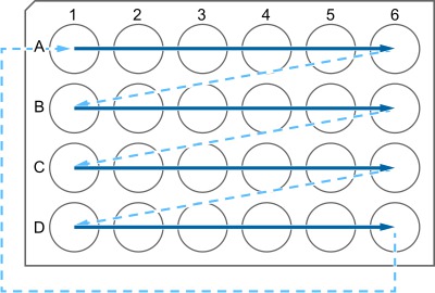
- Cut coronal sections of 40 µm thickness on a freezing microtome (block temperature at -25 to -16 °C). Sequentially transfer sections into antifreeze solution containing wells of a 24-well cell culture plate (see Figure 1). Store at -20 °C.
2. Immunostaining
NOTE: Sections are processed free-floating, usually in 6-well plates equipped with a carrier plate and mesh inserts. As an exception, blocking, antibody incubations, and avidin-biotin complex (ABC) reactions are done in 12- or 24-well plates without mesh inserts (0.5 to 1 ml per well is sufficient, depending on the number of slices that have to be stained). During these steps, transfer sections with the help of a fine brush (rinse with each new solution). All incubations are done with continuous agitation (max 150 rpm).
- Immunohistochemistry (ABC method)
- Transfer sections from antifreeze into tris-buffered saline (TBS) and rinse thoroughly (once O/N at 4 °C, 5 times at room temperature [RT] for 10 min each) to completely remove antifreeze.
- To quench endogenous peroxidase activity, Incubate for 30 min in 1.5% H2O2 in TBS-Tween 20 (TBS-T). Pay attention to bubbling and re-submerge sections if necessary. Rinse 3 times in TBS for 15 min each.
- Optional: Meanwhile, preheat a heating cabinet and 2 N HCl to 37 °C. To denature DNA, incubate sections for 30 min at 37 °C in 2 N HCl. Gently separate sections with the help of a brush.
- Optional: Neutralize sections for 10 min in 0.1 M borate buffer pH 8.5, RT. While transferring, briefly swab the mesh inserts containing the sections on a paper towel to remove HCl leavings. Rinse 2 times in TBS for 15 min each.
- Incubate in TBSplus to permeabilize the tissue and block unspecific antibody binding sites for 1 hr at RT.
- Incubate in primary antibody diluted in TBSplus, O/N at 4 °C. Rinse 3 times in TBS for 15 min each.
- Incubate in biotinylated secondary antibody diluted in TBSplus for 3 hours at RT. Rinse 3 times in TBS for 15 min each. Meanwhile...
- Prepare ABC complex according to manufacturer's protocol (1% A + 1% B in TBS-T). Allow to stand for 30 min at RT before use. Incubate sections in AB reagent for 1 hr at RT. Rinse 3 times in TBS for 15 min each.
- Prepare 50 ml 0.5 mg/ml 3,3′-Diaminobenzidine (DAB) in TBS-T per 6- or 12-well plate, split into two halves, and pipette 4 ml or 2 ml per well, respectively. Transfer sections into DAB solution (CAUTION! DAB is toxic. Follow the specific MSDS provided by the supplier, i.e., wear a lab coat and gloves and use a chemical fume hood).
- Add 0.5 ml 1% H2O2 to the remaining 25 ml DAB solution, mix, and pipette equivalent volumes as above to each well to start the peroxidase reaction. Incubate for 12 min. Rinse 3 times in TBS for 15 min each.
- Mount sections to slides in gelatin and air dry O/N. Coverslip with permanent mounting medium.
- Optional: Counterstain before placing the coverslip.
NOTE: If the signal-to-noise ratio is low because of the high background, repeat the H2O2 treatment after the incubation with AB reagent (step 2.1.8).
- Multiple-immunofluorescence
- Single or simultaneous multiple immunofluorescence
- Transfer sections from antifreeze into TBS and rinse thoroughly (once O/N at 4 °C, 5 times at RT for 10 min each) to remove the antifreeze completely.
- Optional: as steps 2.1.3 - 2.1.4.
- Incubate in TBSplus to permeabilize the tissue and block unspecific antibody binding sites, 1 hr at RT.
- Incubate in primary antibody cocktail (e.g., rat α-BrdU (bromodeoxyuridine), guinea pig α-DCX (doublecortin), goat α-GFP (green fluorescent protein)) diluted in TBSplus, O/N at 4°C. Rinse 3 times in TBS for 15 min each.
- Incubate in a cocktail of fluorochrome-conjugated secondary antibodies (e.g., Rhodamine Red α-rat, Alexa-647 α-guinea pig, Alexa-488 α-goat; all derived in donkey) diluted in TBSplus, 3 hr at RT or O/N at 4 °C. From now on protect sections from light. Rinse 3 times in TBS for 15 min each.
- Mount sections to slides in gelatin and air dry overnight (O/N). Coverslip with aqueous mounting medium.
- Sequential multiple immunofluorescence with primary antibodies from the same host species
- As steps 2.2.1.1 - 2.2.1.3.
- Incubate in first primary antibody (e.g., rabbit α-antigen A), O/N at 4 °C. Rinse 3 times in TBS and once in TBS-T for 10 min each.
- Incubate in first fluorochrome-conjugated secondary antibody (e.g., Rhodamine Red-conj. donkey α-rabbit), 3 hr RT. From now on, protect sections from light. Rinse 3 times in TBS and once in TBS-T for 10 min each.
- Incubate in 10% normal serum from the same host as the primary antibodies (e.g., rabbit serum) for 3 hr RT to saturate open paratopes on the first secondary antibody. Rinse 3 times in TBS and once in TBS-T for 10 min each.
- Incubate in TBSplus with 50 µg/ml unconjugated monovalent Fab fragments directed against the host of the primary antibodies (e.g., α-rabbit IgG (H+L)) to cover epitopes that could be recognized by the second secondary antibody, O/N at 4 °C.
- Rinse at least 3 times in TBS and once in TBS-T for 10 min each. While transferring, briefly swab the mesh inserts containing the sections on a paper towel to remove any Fab leavings.
- Incubate in second primary antibody (e.g., rabbit α-antigen B), O/N at 4 °C. Rinse 3 times in TBS and once in TBS-T for 10 min each.
- Incubate in second fluorochrome-conjugated secondary antibody (e.g., Alexa-488-conj. donkey α-rabbit), 3 hr RT. Rinse 3 times in TBS for 15 min each, mount and coverslip as above.
NOTE: To label antigens with antibodies from different host species, add them to step 2.2.2.2 and the respective secondary antibodies to step 2.2.2.3.