1. Immunolocalization
NOTE: The fluorescent immunolocalization procedure relies on sequential use of two antibodies. The primary antibody is raised against a specific target antigen. The secondary antibody is raised specifically against the primary antibody and for fluorescent techniques is conjugated to a fluorophore (FITC [fluorescein isothiocyanate] in this specific protocol). The primary antibody will be used to detect the target antigen in the sample, and the secondary antibody will be used to mark the location where the primary antibody connected to the sample after washing off the excess of primary antibody. Controls are an important part of this assay and must always be performed to insure the accuracy of the observations. One well on the slide should be reserved for use as a negative control, where the primary antibody treatment will be skipped, and therefore no signal should be observed at the end of the experiment. A positive control must be included in the experiment by treating one well with an antibody which labelling is already known and certain. The positive control is used to confirm the secondary antibody labelling effectiveness and reaction conditions while the negative control tests the secondary antibody specificity.
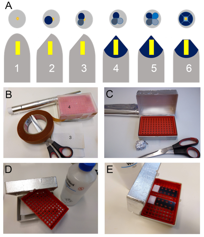
- Prepare an incubation chamber by placing some damped paper towels at the bottom of a pipette tips box and wrapping it with tin foil (Figure 1B-1E). Place the slides in the incubation chamber.
- Pipette a 50 μL drop per 8 mm well of blocking solution (5% nonfat dry milk (w/v) in 1 M phosphate-buffered saline [PBS]) and incubate for 10 min. Remove the blocking solution and wash all wells twice with PBS for 10 min.
- Prepare the primary antibody solutions (see Table 1), 1:5 (v/v) antibody in blocking solution; make an estimate of about 40 μL per 8 mm well.
NOTE: The concentration of the antibody must be adjusted according to the manufacture’s protocol.
- Perform a final wash with double-distilled water (ddH2O) for 5 min, never let the wells dry completely.
- Pipette the primary antibody solution to the reaction wells. Pipette blocking solution to the control wells.
- Close the incubation chamber and let stand for 2 h at room temperature followed by overnight at 4 °C. Prepare the secondary antibody solution, 1% in blocking solution (v/v) about 40 μL per well. Keep it covered with tin foil.
- Wash all wells twice with PBS for 10 min, followed by 10 additional minutes with ddH2O. Make sure that no trace of the blocking solution or deposits is visible on the wells.
- Pipette the secondary antibody solution to all wells. From now on, protect the slides from light.
- Incubate for 3–4 h in the dark at room temperature.
- Wash all wells twice for 10 min with PBS followed by another wash of 10 min with ddH2O.
- Apply a drop of calcofluor (1:10,000 (w/v) fluorescent brighter 28 in PBS) to each well. Without washing, apply a drop of mounting medium (see Table of Materials) to each well and place a coverslip (Figure 2H).
- Observe with a fluorescence microscope, equipped with 10x/0.45, 20x/0.75, 40x/0.95 and 100x/1.40 lens, use UV (for calcofluor stain) and FITC filters, to detect cell wall and immunolocalization respectively (Figure 2I). Use the following wavelengths: excitation/emission (nm) 358/461 for UV and 485/530 for FITC.
- For a better visualization of the results overlap both images with ImageJ or similar.
Table 1: List of useful monoclonal antibodies. The above table represents an example of available antibodies, with information about their targets and where they can be purchased.
| Name | Affinity | Available at |
| LM2 | Arabinogalactan protein | Plant probes (Leeds, U.K) |
| LM5 | Pectins |
| LM6 | Arabinan |
| LM7 | Homogalacturonan |
| LM8 | Xylogalacturonan |
| LM13 | (1-5)-a-L-ARABINAN |
| LM14 | Arabinogalactan protein |
| LM16 | Processed Arabinan - RG-I |
| LM18 | Homogalacturonan |
| LM19 | Homogalacturonan |
| LM20 | Homogalacturonan |
| LM26 | BRANCHED-GALACTAN |
| LM30 | Arabinogalactan protein |
| JIM5 | Low methyl-esterified homogalacturonan | CCRC (Georgia, USA) |
| JIM7 | Highly methyl-esterified homogalacturonan |
| JIM4 | Arabinogalactan protein |
| JIM8 | Arabinogalactan protein |
| JIM13 | Arabinogalactan protein |
| JIM15 | Arabinogalactan protein |
| JIM16 | Arabinogalactan protein |
| JIM20 | Extensin |
| MAC207 | Arabinogalactan protein |