Method Article

Assessing Interneuron Response to Chemoattractive Cues from Periventricular Endothelial Cells

July 8th, 2025

In This Article

Abstract

Loading...
$$\rightleftharpoonup{xx}$$ $$\longleftharp{xx}$$, $$\longrightharp{xx}$$,

Source: Datta, D., et al. Migration, Chemo-Attraction, and Co-Culture Assays for Human Stem Cell-Derived Endothelial Cells and GABAergic Neurons. J. Vis. Exp. (2020).

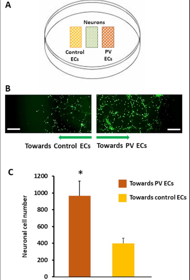
This video shows a method to test interneuron migration in response to chemoattractive cues from periventricular endothelial cells (PVECs). Interneurons are placed in a central compartment, with PVECs and control cells on either side. Increased migration towards PVECs indicates a positive response to their chemoattractive signals.
Protocol

Loading...
$$\rightleftharpoonup{xx}$$ $$\longleftharp{xx}$$, $$\longrightharp{xx}$$,

1. Chemo-attraction Assay

  1. Place a three-well culture insert in the center of a poly-L-ornithine/laminin-coated 35 mm dish using sterile tweezers.
  2. Turn the dish upside down. Mark the boundary around the middle compartment of the insert using a permanent black marker with an ultra-fine tip.
  3. Seed 3 x 10⁴ human GABAergic interneurons in the middle compartment in 70 µL of neuronal medium (Figure 1A).
  4. Seed 104 human periventricular endothelial cells in 70 µL of periventricular endothelial cell medium and 104 control endothelial cells in 70 µL of control endothelial cell medium in the two outer compartments, respectively (Figure 1A).
  5. Add 1 mL of co-culture medium (50% periventricular maintenance medium without GABA and 50% neuronal medium) along the side of the dish to prevent the coating on the dish from drying.
  6. Check under a microscope to verify that cells are not leaking from the insert compartment.
  7. Incubate cells for 24 h at 37 °C and 5% CO2. After 24 h incubation, check under a microscope to verify that cells have attached properly and that there is no leak.
  8. After 48 h of seeding, gently remove the insert using a sterile tweezer. Check under the microscope to verify that the cell layer is not disturbed (day 0).
  9. Remove the medium and add 1 mL of fresh co-culture medium.
    NOTE: Set aside a required number of dishes for day 0 images.
  10. Incubate cells for 36 h at 37 °C and 5% CO2. After 36 h, aspirate medium, fix cells with 4% PFA for 10 min, and wash 3x with 1x PBS.
  11. Stain human GABAergic interneurons with anti-human β-Tubulin or anti-human MAP2 antibody. At the end of the staining procedure, add 1 mL of antifade mounting medium to each dish.

2. Imaging and Data Analysis

  1. Place the immuno-stained assay dish under a microscope at 4X magnification.
  2. Keep one long edge of the rectangular boundary (made in step 5.10 above) in the field of view. Take images of cells in the cell-free space adjacent to that boundary. Acquire images along the right long edge and the left long edge of the rectangular boundary.
    NOTE: Cells positioned diagonally with respect to the rectangle are not considered due to ambiguity in selecting the short- or long-edge as the starting mark. Also, numbers of cells migrating across the short-edge are often significantly fewer (possibly due to lesser number of starting cells along the short-edge) and are not considered.
  3. Open the images in ImageJ. Calculate the distance between each cell and the boundary mark using ImageJ.
  4. To assess migration in terms of cell numbers, set a specific distance from the boundary in the acquired image in ImageJ. Count the number of cells that are present within this distance. Calculate average number, standard devia
  5. tion, and statistical significance using appropriate software.

Access restricted. Please log in or start a trial to view this content.

Results

Loading...
$$\rightleftharpoonup{xx}$$ $$\longleftharp{xx}$$, $$\longrightharp{xx}$$,

Neuronal migration assay: diagram, fluorescent microscopy, bar graph comparing PV and control ECs effects.
Figure 1: Chemo-attraction assay. (A) Schema of the chemo-attraction assay. Using a three-well culture insert, interneurons (IN) were seeded in the middle (green dotted rectangle), while periventricular endothelial cells (PV ECs; orange dotted rec...

Access restricted. Please log in or start a trial to view this content.

Disclosures

Loading...
$$\rightleftharpoonup{xx}$$ $$\longleftharp{xx}$$, $$\longrightharp{xx}$$,
No conflicts of interest declared.

Materials

List of materials used in this article
NameCompanyCatalog NumberComments
Control human endothelial cellsCellular DynamicsR1022
Control endothelial Cells Medium SupplementCellular DynamicsM1019
DMEMF/12 mediumThermofisher Scientific11320033
E6 mediumThermofisher ScientificA1516401
FGF2Thermofisher ScientificPHG0261
FibronectinThermofisher Scientific33016-015
GABASigma-AldrichA2129
HemacytometerSigma-AldrichZ359629
Human GABAergic neuronsCellular DynamicsR1013
Human GABAergic neurons base mediumCellular DynamicsM1010
Human GABAergic neuron Neural supplementCellular DynamicsM1032
LamininSigmaL2020
Mounting MediumVector laboratoriesH-1200
poly-L-ornithinSigmap4957
PBSThermofisher Scientific14190
VEGF-APeprotech100-20
VascuLife VEGF Medium Complete KitLifeline Cell TechnologiesLL-0003Component of control human endothelial cell medium
3-well silicone culture-Insert ibidi80369
35 mm dishCorning430165
4% PFA solutionFisher ScientificAAJ19943K2
Inverted phase contrast microscopeZeissZeiss Axiovert 40C
Fluorescent microscopeOlympusFSX-100
Anti-human β-Tubulin antibodyBiolegend802001
Anti- MAP2 antibodyNeuromicsCH22103

Reprints and Permissions

Request permission to reuse the text or figures of this JoVE article

Request Permission

Tags

Interneuron MigrationChemoattraction AssayPeriventricular Endothelial CellsThree Compartment CultureGABAergic InterneuronsCell Migration AnalysisCo Culture MethodChemoattractive CuesEndothelial Cell ControlPoly L Ornithine Coating

Related Articles