A subscription to JoVE is required to view this content. Sign in or start your free trial.

Method Article

Immunostaining of Neurons Treated with Alpha-Synuclein Aggregates

935 views

July 8th, 2025

In This Article

Abstract

Source: Panattoni, G., et al. Exogenous Administration of Microsomes-associated Alpha-synuclein Aggregates to Primary Neurons As a Powerful Cell Model of Fibrils Formation. J. Vis. Exp. (2018)

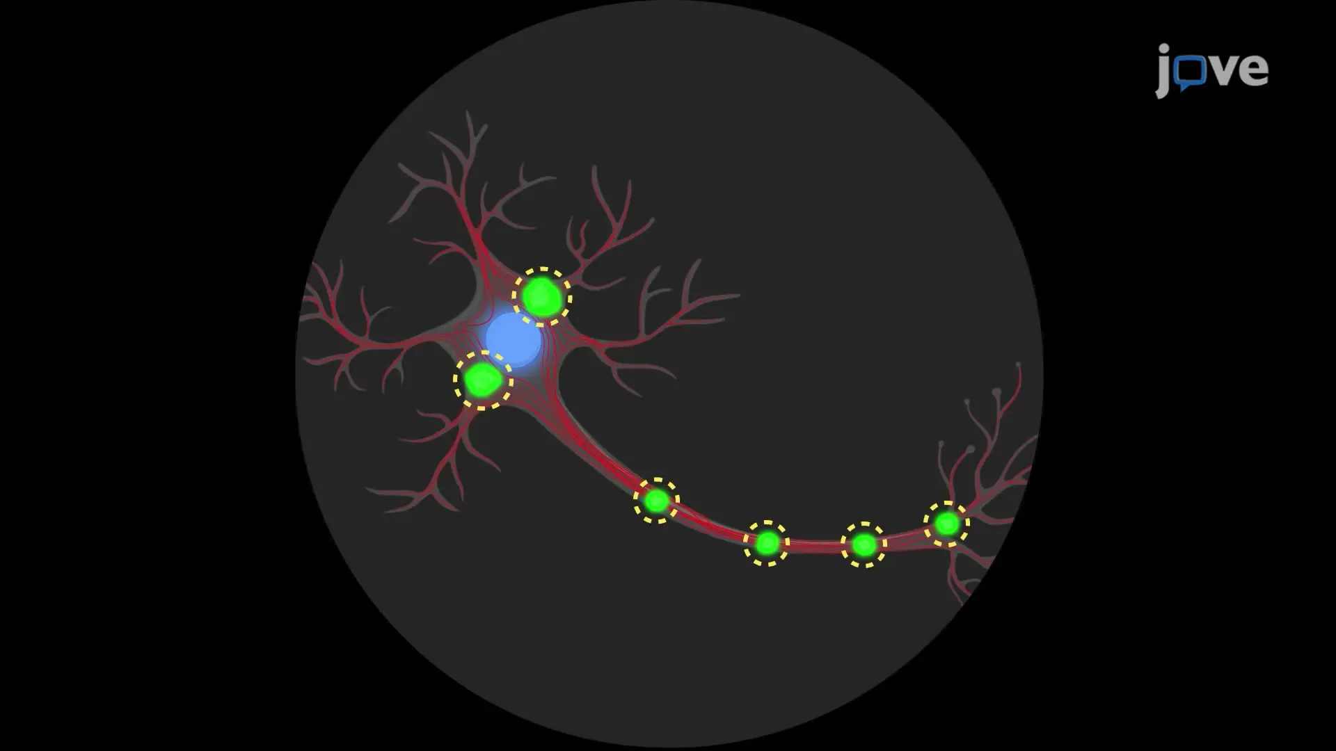
This video demonstrates the immunostaining of neurons treated with alpha-synuclein (αS) aggregates. The neurons are first treated with αS aggregates. They are then fixed, permeabilized, and treated with a blocking solution. Primary antibodies are added, followed by a fluorescently labeled secondary antibodies and a nuclear stain. The sample is then mounted and observed under a microscope.

Protocol

1. Neurons Treatment

Note: The treatment has been performed at Division (DIV) 7. All the steps are carried on under a cell culture hood in sterile conditions. An example of cortical neuronal culture density at DIV 7 is illustrated in Figure 1.

  1. Pool microsomes-associated αS aggregates obtained from the spinal cord of three different diseased Transgenic mice to have a 1 µg/µL solution, using the original homogenization buffer [composed of 250 mM sucrose, 20 mM HEPES (4-(2-hydroxyethyl)-1-piperazineethanesulfonic acid), 10 mM KCl (Potassium Chloride), 1.5 mM MgCl2 ( (Magnesium Chloride), 2 mM EDTA (Ethylenediaminetetraacetic acid), and 1x phosphatase/protease-inhibitors] for the dilution.
  2. Take a neuron culture on poly-D-Lysine coverslips in 24 well plates containing medium (2% B27, 10x gentamicin, and 2 mM glutamine). Remove 1/3 of the medium and replace it gently with the fresh medium containing 2% B27, 1x gentamicin, and 2 mM glutamine.
  3. Add 1 µg of pooled microsomes-associated alpha-synuclein (αS) aggregates to the cell medium. Return neurons to the incubator at 37 °C.
  4. Every 3 days for 1 week, add 1/3 of fresh medium. Do not replace the medium. Just add it.
  5. After 1 week of treatment, remove 1/3 of the medium and replace it gently with the fresh medium containing 2% B27, 10 µg/mL gentamicin, and 2 mM glutamine. Repeat every 3 days.
  6. Fix neurons after 2 weeks of treatment (DIV 21).

2. Immunofluorescence

  1. Fix neurons with 2% paraformaldehyde in Phosphate-Buffered Saline (PBS) 1x and 5% sucrose solution for 15 min at room temperature (RT) without shaking under a chemical fume hood.
  2. Remove the fixing solution. Briefly wash with PBS 1x, 3 times.
    NOTE: Perform all the washing steps gently because primary neurons do not stick firmly to poly-lysine coverslips.
  3. Permeabilize neurons with 0.3% of non-ionic surfactant in PBS 1x for 5 min at RT.
  4. Briefly wash with PBS 1x, 3 times.
  5. Incubate neurons with 3% Fetal Bovine Serum (FBS) in PBS 1x for 30 min at RT to block unspecific binding sites on an orbital shaker.
  6. Incubate neurons with appropriate primary antibody dissolved in 3% FBS in PBS 1x, O/N at 4 °C on an orbital shaker.
    NOTE: syn303 (1:1,000), mouse αS (1:200), pser129-αS (1:1,000), and Tau (1:10,000) antibodies were used.
  7. Remove the antibody solution and wash, briefly, with PBS 1x, 3 times.
  8. Incubate neurons with appropriate fluorescent secondary antibody dissolved in 3% FBS in PBS for 1 h at RT in the dark on an orbital shaker.
  9. Remove the antibody solution and wash, briefly, with PBS 1x, 3 times.
  10. Stain neurons with DAPI (4′,6-diamidino-2-phenylindole) solution (0.1 µg/mL in PBS 1x) for 15 min at RT in the dark on an orbital shaker.
  11. Remove the antibody solution and wash, briefly, with PBS 1x, 3 times.
  12. Mount coverslips on a slide using an antifade mounting medium.

Access restricted. Please log in or start a trial to view this content.

Results

Cell culture under microscope; neuron-like network; biological research; microscopy study.
Figure 1. Cortical neuronal cultures. Representative image showing density at DIV 7 of cortical neuronal cultures. Images were taken with an inverted light microscope, 10X objective. Scale bar = 100 µm.

Access restricted. Please log in or start a trial to view this content.

Disclosures

No conflicts of interest declared.

Materials

List of materials used in this article
NameCompanyCatalog NumberComments
SucroseSigma-Aldrich84097-1KG
HepesSigma-AldrichH0887-100ML1M pH=7-7.6
EDTASigma-Aldrich0390-100mlpH=8 0.5M
Magnesium chlorideSigma-AldrichM8266-100G
Sodium carbonateSigma-AldrichS7795-500G
Sodium bicarbonateSigma-AldrichS5761-500G
MethanolSigma-Aldrich322415-6X1L
KClSigma-AldrichP9541-500G
cOmplete MiniRoche11836170001protease inhibitor
PhosStopRoche4906837001phosphatase inhibitor
BCA Protein Assay KitEurocloneEMPO14500
Criterion TGX 4-20% Stain Free, 18 wellsBiorad5678094
Supported Nitrocellulose membraneBiorad16200970.2 μm
Blotting-Grade BlockerBiorad1706404Non-fat dry milk
SuperSignal West Pico Chemiluminescent SubstrateTermo Fisher Scientific34077
Nitric acidSigma-Aldrich100441100065%
Glass CoverslipsTermo Fisher Scientific1014355118NR118 mm x
Poly-D-LysineSigma-AldrichP7280
Hank's Balanced Salt SolutionTermo Fisher Scientific14170-500 mL
Penicillin/StreptomycinTermo Fisher Scientific1514012210,000 U/mL, 100 mL
Dulbecco's Modified Eagle's MediumTermo Fisher ScientificD5796-500 mL
Trypsin-EDTATermo Fisher Scientific154000540.50%
B27 SupplementTermo Fisher Scientific1750404450X
GlutamaxTermo Fisher Scientific35050-038100x
DNAseSigma-AldrichD5025
Fetal bovine serumEurocloneEC50182L
GlutamateSigma-Aldrich1446600-1G
GentamicinTermo Fisher Scientific1571010 mg/ml
Neurobasal MediumTermo Fisher Scientific10888-022
Cytosine arabinoside (AraC)Sigma-AldrichC3350000
VECTASHIELD antifade mounting mediumVector LaboratoriesH-1000
DAPITermo Fisher Scientific62247
90 Ti rotorBeckmanN/AUltracentrifuge rotor
Optima L-90K UltracentrifugeBeckmanN/A
Syn-1 antibody, clone 42BD Biosciences610786anti-mouse WB: 1:5000
Syn303 antibodyBioLegend824301anti-mouse IF: 1:1000
Tau antibodySynaptic Systems314 002anti-rabbit IF: 1:10,000
pser129-αS antibodyA gift from Fujiwara et al, reference 19anti-rabbit WB: 1:5000
pser129-αS antibodyAbcamab51253anti-rabbit IF: 1:1000
Mouse αS (D37A6) XPCell Signaling4179anti-rabbit IF 1:200
Alexa fluor 555-conjugated anti-rabbit antibodyTermo Fisher ScientificA27039
Alexa fluor 488-conjugated anti-mouse antibodyTermo Fisher ScientificA-11029
Microson XL-2000MisonixSonicator
Ultra Bottles (Oakridge Bottles), PCB, 16x76mm, Assembly, Noryl Cap, Beckman-typeScience Service EUS4484Ultracentrifuge tubes
AXIO Observer Inverted Light MicroscopeZeissN/A
TCS SP2 laser scanning confocal microscopeLeicaN/A
Inverted epi-fluorescence microscopeNikonN/A
Triton x-100Sigma-AldrichX100-500MLNonionic surfactant

Tags

Neuron ImmunostainingPrimary Antibody IncubationFluorescent Secondary AntibodyDAPI Nuclear StainingParaformaldehyde FixationMembrane PermeabilizationBlocking Solution TreatmentFluorescence Microscopy ObservationCoverslip Mounting Procedure