1. Establishing the BBB Model
Note: For establishing the blood-brain barrier (BBB) model we suggest using commercially available primary rat brain microvascular endothelial cells (RBMECs) and rat cortical astrocytes (RCAs). All steps must be performed with sterile reagents and disposables handled in a laminar flow hood.
- Cell Culture
- Coat cell culture flasks with poly-L-lysine 100 µg/ml (1 hr at room temperature, RT) or fibronectin 50 µg/ml (1 hr at 37 °C) to promote the attachment of RCAs or RBMECs, respectively. Then, thaw 1 x 10⁶ RCAs and 5 x 10⁵ RBMECs in endothelial cell medium, supplemented with 5% fetal bovine serum, 1% endothelial cell growth supplement, and 1% penicillin/streptomycin (sECM). Seed RCAs in a T175 flask in 20 ml sECM and RBMECs in a T75 flask in 10 ml sECM.
Note: The thawing and seeding passages of RCAs and RBMECs must be adjusted in terms of cell density and time in culture according to the number of experimental conditions to be tested with the BBB model. By starting with 1 vial of 1 x 10⁶ RCAs and 1 vial of 5 x 10⁵ RBMECs, it is possible to obtain up to 20 BBB-bearing inserts.
- Maintain the cells at 37 °C and 5% CO2 in a humidified atmosphere for approximately 6 days until about 80% confluence for RCAs and over 90% confluence for RBMECs. Detach cells using trypsin-ethylenediamine tetraacetic acid (trypsin-EDTA) solution (trypsin 1:250) for 5 min (1 ml trypsin for T75 flasks and 2 ml for T175 flasks). Stop trypsin activity with sECM (2:1), centrifuge cell suspensions at 750 × g for 5 min, and resuspend the pellets in 60 ml sECM.
- Split the RBMECs into 3 T175 flasks and culture in sECM for another 3 days before seeding onto inserts. Count the total number of living RCAs by observing a cell suspension diluted 1:1 with trypan blue under an optical microscope in a Burker chamber.
- Cell Seeding onto Inserts
- Treat one side of the polyethylene terephthalate (PET) membrane of 6 multi-well transparent inserts with poly-L-lysine 100 µg/mL and the other side with fibronectin 50 µg/mL, to allow attachment of RCAs and BMECs. Handle the inserts with tweezers in order to avoid contact with the PET membrane.
- Place the inserts into a 6 well plate and add fibronectin solution (minimum 500 µl) into the upper chamber. After 1 hr of incubation at 37 °C, remove the fibronectin solution, take the inserts off the multi-well plate and put them upside down on the bottom of a 150 cm2 Petri dish, which is used here as a sterile support for the already coated inserts.
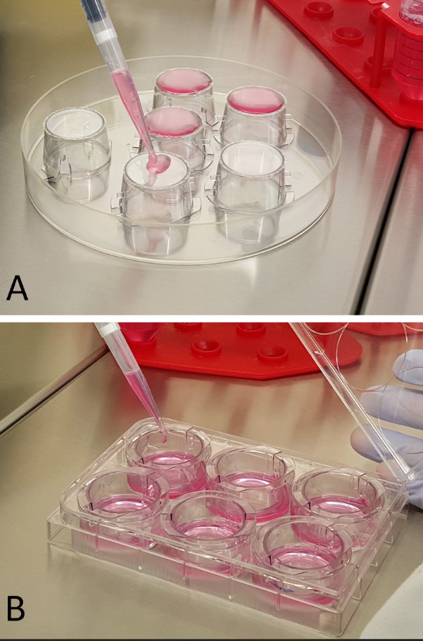
- Gently add 800 µl of poly-L-lysine onto the bottom side of the insert (as shown in Figure 1A; referred to as RCAs seeding), and keep the solution on the inserts for 1 hr at RT.
- Aspirate the solution and let the inserts dry at RT for 15-30 min. The inserts are now ready for cell seeding but can also be stored in the multi-well plate for several days at 4 °C before proceeding with the BBB construction.
Note: Remember to keep at least 3 coated inserts free from cells to be used as controls to validate BBB establishment by FD40 flux measurements.
- Seed RCAs (35,000/cm2) onto the bottom side of the poly-L-lysine-coated inserts by dropping 800 µl of cell suspension onto the upside-down insert (Figure 1A). Leave the RCA suspension on the inserts for 4 hr at RT in order to allow efficient attachment of the cells to the membrane.
- Aspirate the residual solution, place the inserts into wells containing 2 ml of sECM, and maintain the multi-well plate at 37 °C and 5% CO2 in a humidified atmosphere, changing the sECM every 2 days.
- After 3 days, when the RCAs have coated the lower face of the insert, seed RBMECs onto the upper side of the insert, following these steps:
- Detach RBMECs from T175 flasks and count the total living cells, as reported in steps 1.1.2 and 1.1.3.
- Seed RBMECs (60,000/cm2) onto the upper surface of the insert in sECM (1,000 µl) (Figure 1B), leaving 3 inserts with the astrocytes only, to be used as TransEndothelial Electrical Resistance (TEER) background. Place the multi-well plate in standard culture conditions. Maintain the system to culture for at least 3 days, changing the sECM in the inner and lower chamber every 2 days.
2. BBB Validation
- TEER Measurement
- On the 3rd day of co-culture, check the TEER by inserting the BBB-bearing inserts into an appropriate chamber (with a cap and where both chamber and cap contain a pair of voltage-sensing and current electrodes), filled with 4 ml of sECM. Connect to an epithelial tissue volt/ohmmeter.
- Together with the TEER measurements of the cell BBB systems, record the TEER values of the 3 inserts bearing the RCAs that will be subtracted from the values obtained with the BBBs. Multiply the resulting TEER values by the surface of the insert (4.2 cm2) in order to express the results as Ω x cm2.
- From the 3rd day of co-culture, measure the TEER every day. Note: After an initial period where the TEER increases, the recorded values should remain stable for at least two consecutive days (usually between the 5th and the 7th day of co-culture). At that point, the BBB is ready for the second step of validation (section 2.2) and/or for the trans-BBB experiments.
Note: The procedure described in section 2.1 can be performed on the same BBB-systems devoted to the following permeability experiments (section 3). Operate under sterile conditions.
3. Trans-BBB Permeation of FITC-loaded Ferritins (FnN)
Note: A recombinant variant of human ferritin (Fn), produced in Escherichia coli and assembled in nanocages (FnN). FnN are loaded with fluorescein isothiocyanate (FITC), and the concentrations of both Fn and the loaded molecules are accurately determined.
- Trans-BBB Flux of FITC and FITC-FnN
- Measure the FITC-FnN flux from the upper to the lower chamber of validated BBB models (usually at the 5th - 7th day of co-culture), compared to that of the free dye after 7 and 24 hr of incubation, according to the following steps:
- Add FITC-FnN (50 µg/ml FnN, 1.1 µM FITC) or an equal amount of free FITC into the upper chamber of at least 6 BBB systems for each formulation, in order to withdraw 2 ml of sECM solution from the lower chamber of at least 3 inserts after 7 hr, and from the lower chamber of other 3 inserts after 24 hr.
- Measure the fluorescence intensity of 500 µl of the collected samples by spectrofluorometer (λex 488 nm, λem 515 nm, slit 5).
- Obtain the sECM background fluorescence by measuring 500 µl of medium using the same analytical parameters. Subtract the sECM background fluorescence from all FITC or FITC-FnN fluorescence values.
- Determine the concentration of permeated FITC or FITC-FnN by comparing the obtained fluorescence values with two different calibration curves produced with known concentrations (i.e. 6.87, 13.75, 27.5, 55 nM) of the free or nanoformulated dye dissolved in 500 µl sECM.