All procedures involving animal samples have been reviewed and approved by the appropriate animal ethical review committee.
1. Staining of the Primary Antibody and Biocytin
(Day 1)
NOTE: The following immunostaining procedure is for free-floating sections and requires continuous shaking at a low speed (2 revolutions/min, rpm) on a shaker for all incubation steps.
- Wash the tissue 3x for 10 min each in 0.1 M PBS (pH = 7.4).
NOTE: 0.1 M PBS can be commercially purchased or made in the laboratory as follows (makes 1 L): 8 g of NaCl, 0.2 g of KCl, 1.44 g of Na2HPO4, and 0.24 g of KH2PO4 in 1 L of dH2O.
NOTE: If the desired neurochemical marker is known, staining for a protein/peptide can be performed simultaneously with biocytin staining (steps 1.2 - 1.4). If staining for biocytin only, proceed to step 1.5.
- OPTIONAL: Block with 10% Blocking Serum (BS; normal goat serum (NGS) diluted in 0.3% Triton X-100 in PBS for 1 h at RT).
NOTE: Each well should receive the same volume of the solution; the recommended volume is between 250 - 500 µL/well. The selection of the blocking serum is based on the species in which the secondary antibodies are raised (e.g., NGS is used for dye-conjugated goat anti-(respective primary antibody species)). If the need arises to use secondary antibodies raised in different species to immunostain sections, include 10% of each normal serum in blocking step (2) and 3% of each normal serum for the primary and secondary antibody incubation steps.
- (OPTIONAL) Incubate the sections in primary antibody, CB1R (polyclonal, guinea pig), with 0.3% Triton X-100 and 3% BS in 0.1 M PBS at RT O/N. CB1R is used to identify cannabinoid receptor type 1 expression.
NOTE: This step is optional and not required to reveal the biocytin filling. Figure 2 illustrates a section labeled for biocytin and CB1R during the first staining process. O/N incubation at RT is sufficient for most of the primary antibodies; however, certain primary antibodies, including CB1R, require 3 - 5 d of incubation. If the incubation needs to be longer than 1 d, it is recommended to incubate at 4 °C because Triton X-100 can permeabilize slices and can lead to the disintegration of the tissue during prolonged incubation at room temperature. Include a negative control lacking a primary antibody in at least one section for each series of experiments as a control for the specificity of the secondary antibody. For negative controls, the primary antibody is omitted, but the 0.3% Triton X-100 in 0.1 M PBS and BS are added.
(Day 2)
- Wash the brain sections 3x for 10 min each in 0.1 M PBS (pH = 7.4).
- Incubate the sections in streptavidin red dye conjugate (concentration: 1:1,000; excitation: 594 nm with emission in the visible red spectrum) with 0.3% Triton X-100 and 3% BS in 0.1 M PBS at 4 °C O/N in the dark (wrapped in aluminum foil) to reveal the biocytin. OPTIONAL: Include a secondary antibody, goat anti-guinea pig (concentration: 1:500; excitation: 488 nm and emission in green), with the above incubation following the optional primary incubation in step 1.3.
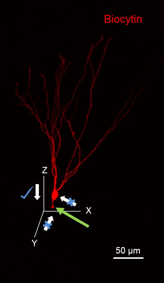
NOTE: A secondary antibody will be needed only if the optional primary antibody staining (step 1.3) is performed. To visualize biocytin, a streptavidin-fluorophore conjugate with an emission spectrum in red is recommended, as it helps visualize finer axons in detail and with better contrast (Figures 1 - 3).
(Day 3)
- Wash brain sections 3x for 10 min each in 0.1 M PBS (pH = 7.4).
- To protect the fluorescence and to facilitate re-staining in the future, mount the sections in an aqueous-based mounting medium and seal the edges of the coverslip with clear nail polish.
NOTE: It is best to restain sections as early as possible to avoid difficulties removing the nail polish from the slide, mold infestation, and tissue dehydration due to the mounting medium leaking from the glass slide. Microbial contamination can be prevented by using 0.02% sodium azide in PBS.
CAUTION: Sodium azide is highly toxic and flammable when it is in contact with water. Please refer to standard operating procedures for the handling and disposal of hazardous chemicals.
(Days 4 - 7)
- Perform confocal imaging with excitation at 594 and 488 nm and at a magnification of 20X or 40X to reveal biocytin-filled neurons in red and neurochemical markers (CB1R) in green. Assess colabeling (Figure 2).
- Set the camera exposure to less than 100 ms to avoid photobleaching and obtain confocal image stacks of the axonal and dendritic arbors of the entire neuron. Use confocal image stacks and neuron tracing software packages to reconstruct the neuronal morphology.