All procedures involving animal models have been reviewed by the local institutional animal care committee and the JoVE veterinary review board.
1. Bilateral renal denervation
- Select the mice with elevated blood pressure (BP) ≥140/90mmHg or 25% increase in systolic BP/diastolic BP, 1 week after the Ang II infusion.
- Record animal weight before surgery and choose animals with a minimum weight of 24 g for renal denervation surgery.
- Anesthetize the mice using sodium pentobarbital. Confirm anesthesia depth with a negative toe pinch reflex.
- Remove the hair on the abdomen with a shaver. Perform this procedure carefully and thoroughly to avoid any surgical contamination.
- Place the mice on the operating table, keeping the abdomen up, and fix its limbs with tape. Disinfect the abdominal skin with povidone-iodine, followed by three wipes with 70% ethanol.
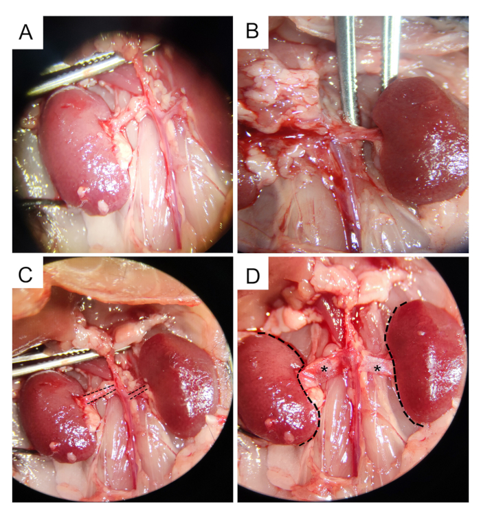
- Make a 2 cm ventral midline abdominal incision using a scalpel blade. Pull back the intestine with gauze soaked in 37 °C saline to expose the left renal artery. Carefully yet bluntly dissect the fat away from the renal artery using curved tweezers. (Figure 1A-C).
- Cut the weighing paper into a rectangle the same size as the renal artery with sterile sharp scissors. For reference, cut the weighing paper in the same size as shown by the dotted line in Figure 1C.
NOTE: It is a critical part of the surgery to try to cut several pieces of the weighing paper at a time to keep the same shape.
- Dip the weighing paper in a 10% phenol/ethanol solution for at least 30 s. Cover the surface of the left renal artery and wrap the vessel with the weighing paper, keeping it for 2 min (Figure 1D). Use gauze to protect the surrounding tissues to prevent the weighing paper from touching the surrounding kidney and intestine.
NOTE: The phenol solution is stable in plastic tubes but not glass vials. Therefore, the solution must be freshly prepared for every experiment.
- Perform the same procedure for the right renal artery. Perform the sham surgery with weighing paper immersed in saline.
- Reposition muscles into their initial position and close the peritoneum with 6-0 Vicryl sutures in an interrupted suture. Then, close the skin with interrupted 4-0 nylon sutures. Monitor all mice until fully recovered.
2. Post-operative care
- Apply povidone-iodine to the incision and place the animal in a warmed electric blanket for recovery and post-operative monitoring.
- Monitor the mice twice a day to assess for redness, swelling, and pain or abdominal infection. Provide meloxicam (0.5 mg/kg, SC) to all mice about 1 h before and 24 h after the RDN procedure.