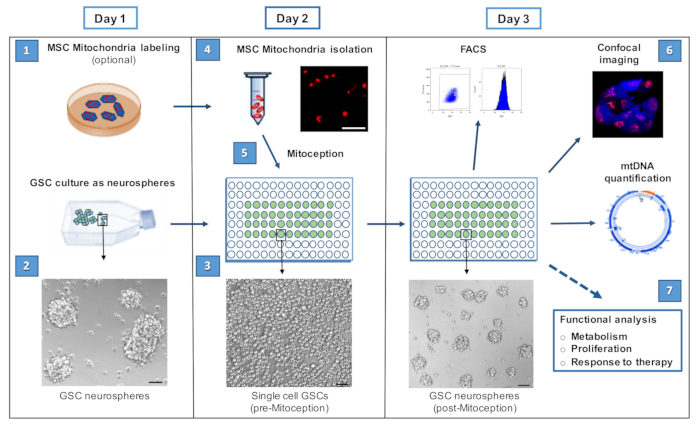
Day 1
1. Labeling of the Mesenchymal Stem Cell (MSC) Mitochondria
- Two days before the mitochondria preparation, seed human MSCs in a 100 mm cultAure dish, in 10 ml (Minimum Essential Medium Eagle)/FBS (Fetal bovine serum) 10%, so as to have 4 x 10⁵ MSCs in culture on Day 1.
- Rinse MSCs with PBS (4 ml) and add 4 ml αMEM/FBS 1% (prewarmed to 37 °C).
- Add the required amount of mitochondria vital dye and incubate cells for 30 min in the 37 °C incubator.
- Remove the mitochondria dye solution, rinse cells twice with 4 ml prewarmed (37 °C) αMEM/FBS 1% and add back 4 ml αMEM/FBS 10%. Incubate cells at 37 °C.
- Change the culture medium (10 ml αMEM/FBS 10%) after 30 min and, another time, 2 hr later.
Day 1
2. Labeling of the Glioblastoma Stem Cells (GSC)
- Dissociate GSCs Gb4 cell line (10 x 106 cells) grown as neurospheres on poly-HEMA coated cell culture flasks
- Seed GSCs in a 48-well plate at 105 cells/well in GSC proliferation medium (500 µl) (see Table 1).
- Centrifuge the plate at 270 x g for 5 min at 20 °C.
- Add the required amount of cell vital dye and incubate for 30 min at 37 °C.
- Add 500 µl of GSC basal medium (Table 1) per well of the 48-well plate.
- Centrifuge the plate at 270 x g at 20 °C for 5 min. Aspirate the supernatant.
- Repeat steps 2.5 to 2.6.
- Add 500 µl of GSC basal medium and incubate at 37 °C for 30 min.
- Centrifuge the plate at 270 x g at 20 °C for 5 min. Aspirate the supernatant.
- Add 500 µl of GSC proliferation medium per well of the 48-well plate and incubate in the 37 °C incubator.
Note: The amount of GSCs indicated (10 x 106 cells) allows performing the different dose-response experiments and FACS controls. Once the experimental conditions are more precisely defined this amount can be scaled down.
3. Seeding of Glioblastoma Stem Cells
- Collect the GSC (Glioblastoma stem cells ) neurospheres (10 x 106 cells) by centrifugation at 270 x g for 5 min, at 20 °C in a 50 ml tube.
- Wash cells with 5 ml of HBSS (Hanks' Balanced Salt Solution), and centrifuge at 270 x g for 5 min at 20 °C.
- Aspirate the supernatant.
- Gently resuspend the GSC pellet in 100 µl trypsin-EDTA(Ethylenediamine tetraacetic acid) (0.25%) (per 10 x 106 cells)
- Incubate at 37 °C for 3 min.
- Add 10 µl CaCl2 (20 mM) and 2 µl DNase I (10 mg/ml).
- Dissociate the neurospheres by gently pipetting up and down (30-50x) with a P200 pipette. Avoid bubbles. Check under the microscope that all GSCs are dissociated.
- Add 10 µl trypsin inhibitor (5%) and 10 ml HBSS.
- Centrifuge GSCs at 270 x g for 7 min at 20 °C.
- Discard the supernatant and add 10 ml GSC basal medium .
- Count GSCs with a Thoma counting chamber and then centrifuge the cells at 270 x g for 7 min at 20 °C.
- Add the appropriate volume of GSC proliferation medium to reach the cellular concentration of 106 GSCs/ml.
- Seed 105 GSCs (100 µl of the cell suspension) per well of a 96-well plate.
- Centrifuge the plates at 270 x g for 7 min at 20 °C to get GSCs at the bottom of wells.
- Incubate the 96-well plate at 37 °C.
Day 2
4. MSC (Mesenchymal stem cells) Mitochondria Isolation
- Adjust the microtube centrifuge temperature to 4 °C.
- Prepare two 1.5 ml tubes "A" and "C" containing the reagents for the mitochondria extraction (200 µl reagent A and 400 µl reagent C, both containing the EDTA-free protease inhibitors). Prepare 2 other tubes, labeled "MSC" and "Mito". Keep all tubes on ice. Also prepare tubes for the mitochondria serial dilutions.
- Wash MSCs with 10 ml prewarmed (37 °C) PBS (Phosphate buffered saline).
- Wash MSCs with 2 ml trypsin (no EDTA) for 10 sec and add 1 ml trypsin (no EDTA). Incubate cells for 5-10 min at 37 °C.
- Recover the MSCs by adding 10 ml αMEM /FBS 10%, transfer to a 50 ml tube.
- Centrifuge cells at 270 x g for 5 min at 20 °C.
- Discard the supernatant and add 10 ml αMEM/FBS 10% to the cell pellet.
- Count the MSCs with a Malassez counting chamber.
- Centrifuge MSCs (4-5 x 10⁵) at 270 x g for 5 min at 20 °C.
- Discard the supernatant, add 1 m ice-cold αMEM/FBS 10% to the cell pellet and transfer the cells to the "MSC"-labeled tube. Keep the tube on ice.
- Centrifuge the tube containing the MSCs at 900 x g for 5 min, at 4 °C.
- Remove all residual medium from the tube.
- Add 200 µl of Mitochondria Isolation Reagent A (containing the EDTA-free protease inhibitors). Vortex at medium speed for 5 sec and leave tubes on ice for exactly 2 min.
- Add 2.5 µl of Mitochondria Isolation Reagent B. Vortex at maximum speed for 10 sec, and then leave tubes on ice. Repeat every 30 sec for 5 min.
- Add 200 µl of Mitochondria Isolation Reagent C (containing the EDTA-free protease inhibitors). Mix by tilting the tube (roughly 30 times, do not vortex). Centrifuge the tube at 700 x g for 10 min at 4 °C.
- Transfer the supernatant (containing the MSC mitochondria) to the "Mito" tube.
- Centrifuge at 3,000 x g, 15 min at 4 °C, to get the mitochondria pellet. Discard the supernatant. The pellet contains the isolated mitochondria.
- Rinse the mitochondria pellet with 200 µl of the Reagent C. Then, centrifuge at 12,000 x g, 5 min at 4 °C, to get the mitochondria pellet.
5. Transfer of Isolated MSC Mitochondria to GSCs (MitoCeption)
- Add 200 µl of the pre-cooled (0 °C) GSC proliferation medium to the mitochondria pellet isolated from MSCs (4 x 10⁵).
- Dilute the mitochondria preparation (in GSC proliferation medium) to consistently add 20 µl of mitochondrial suspension to the GSCs.
- Add the volume of isolated mitochondria into the wells of the 96-well plate containing the GSCs , at the desired concentration (0.1 to 10 µg). Add the mitochondria slowly, close to the bottom of the well, covering the entire surface at least once.
- Centrifuge the 96-well plate containing the mitochondria recipient GSCs with the MSC mitochondria and the control plate at 1,500 x g for 15 min at 4 °C.
- Place the culture plates in the 37 °C cell incubator immediately after centrifugation.
Note: The mitochondria transfer protocol relies on the centrifugation of the mitochondria suspension on the cultured cells at the adequate centrifugation force, with a number of centrifugations that can be adjusted depending on the system of mitochondria donor/recipient cells.
Table 1: Culture Media.
| Cell culture medium | Composition |
| GSC basal medium | DMEM/F-12 supplemented with |
| Insulin 20 mg/ml |
| N2 supplement 1x |
| Glucose 3 g/L |
| L-glutamine 2 mM |
| GSC proliferation medium | Add to the basal medium: |
| B27 supplement 1x |
| EGF 10 ng/ml |
| bFGF 10 ng/ml |
| Fungine 10 mg/ml |
| Fungizone 0.25 mg/ml |
| Heparin 2 mg /ml |
| Ciprofloxacin 2 μg / ml |
| Gentamicin 2 μg/ml |
| MSC proliferation medium | αMEM supplemented with |
| L-glutamine 2 mM |
| 10% FBS |
| bFGF 2 ng/ml |