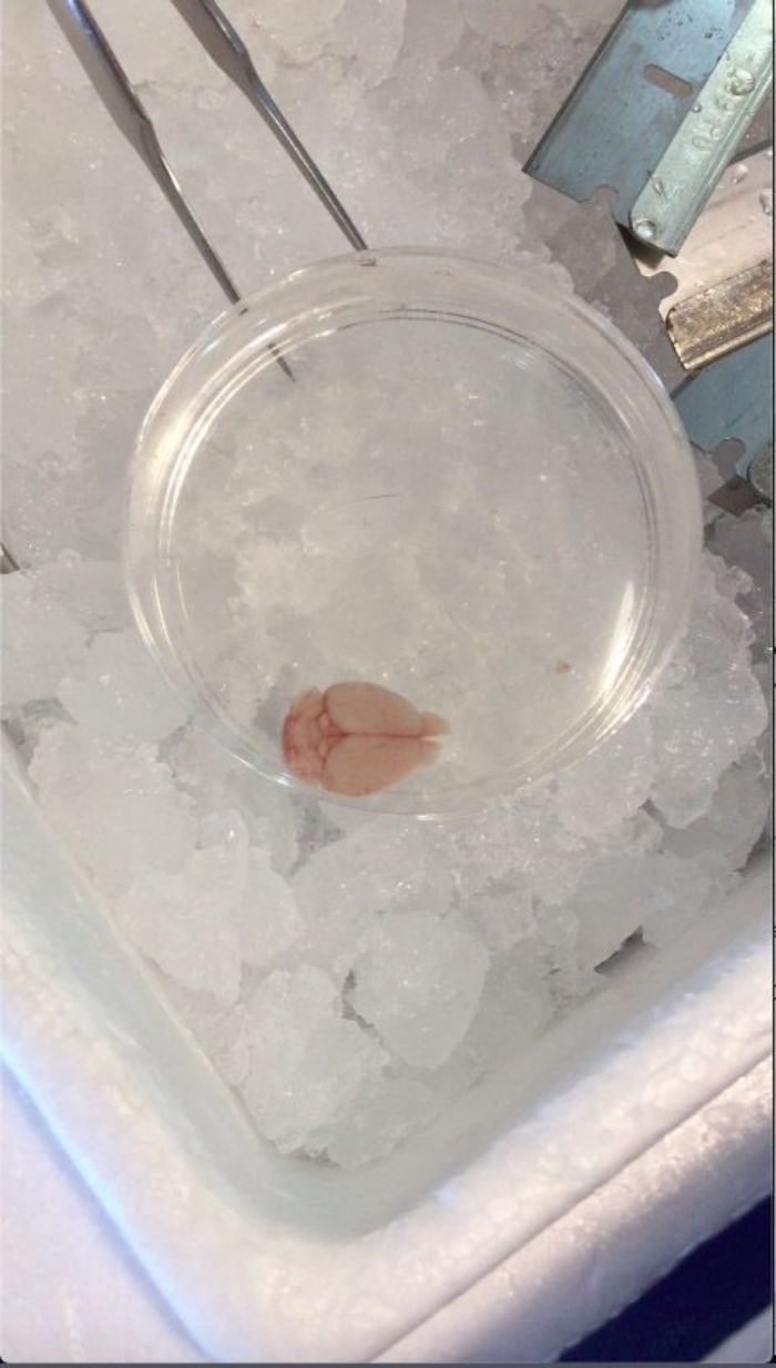
All procedures involving animal samples have been reviewed and approved by the appropriate animal ethical review committee.
1. Preparation of the working solutions and buffers
NOTE: On the morning of the assay, prepare the following solutions. This calculation is based on the necessary solutions to process two brain regions (i.e., the prefrontal cortex [PFC] and hippocampus [HPC]) from 12 mice.
- Prepare artificial cerebrospinal fluid (aCSF, pH = 7.4) as mentioned in Table 1. Dispense 750 µL of aCSF into each sampling tube, and place them in the metal temperature block on ice to pre-chill the buffer.
- Prepare the lysis buffer as mentioned in Table 2. Store on ice (400 µL to be used per sample).
- Prepare a 52 mM Bis(sulfosuccinimidyl)suberate or BS3 stock solution (26x) in 5 mM sodium citrate buffer (pH = 5.0).
- First, prepare 100 mM citric acid (stock A) and 100 mM sodium citrate (stock B).
- Dilute stock A and stock B at a ratio of 1:20 with deionized water. Add 100 µL each to 1.9 mL of water to prepare 5 mM citric acid (stock C) and 5 mM sodium citrate (stock D), respectively.
- Mix 410 µL of stock C and 590 µL of stock D to prepare a 5 mM sodium citrate buffer (pH = 5.0) (solution E, 1 mL).
- Confirm the pH of solution E using a pH indicator strip.
- Dissolve 24 mg of BS3 in 806.4 µL of solution E by vortexing for 30 s to prepare the BS3 stock solution (26x).
- Prepare another 1 mL of solution E to use as vehicle control for the non-crosslinking samples.
NOTE: Prepare BS3 stock solution when everything else is ready, and the experiment is about to begin. The BS3 should be stored desiccated at 4 °C until use. Once reconstituted, BS3 remains active only for approximately ≤3 h. As the pH of the 5 mM sodium citrate buffer (solution E) is reported to rise over time, causing accelerated BS3 hydrolysis, it is recommended that solution E is prepared fresh from stock solutions A and B. Due to the limited solubility of BS3 at low temperatures, the reconstituted BS3 should be kept at RT. Use the reconstituted BS3 within 3 h, and do not freeze/thaw or reuse the reconstituted BS3.
2. Dissection of brain tissues
NOTE: From this step on, at least two people should work together in a coordinated manner. While one person focuses on the animal dissection, the other person should work as a timekeeper and help coordinate the assay.
- Bring the first animal for dissection from the housing area to the dissection room.
NOTE: As acute stressors (e.g., a novel environment, the smell of blood) may affect the brain protein dynamics, the animals should be kept in their home cages placed far from the dissection area and then be brought individually into the dissection room for immediate decapitation.
- Euthanize the mouse by cervical dislocation followed by decapitation. Remove the brain rapidly out of the skull and submerge it in ice-cold PBS in a Petri dish for 10-15 s (Figure 1).
NOTE: The animals are not anesthetized for BS3 experiments since any anesthetic agent could potentially influence the surface presentation level of the neurotransmitter receptors.
- Place the chilled brain into the brain matrix on ice, with the ventral side of the brain facing up (Figure 2).
- Insert the first razor blade through the border between the olfactory bulb and the olfactory peduncle to cut the brain coronally (Figure 3). Using three to four additional razor blades, serially cut the anterior part of the brain coronally with 1 mm intervals.
- Lift the coronal slices off the brain matrix by holding all the inserted razor blades together, leaving the posterior part of the brain behind in the brain matrix. Use forceps to separate the razor blades from one another, and place them on the flat, chilled surface with the brain slice facing up (Figure 4).
- Identify the slices containing the region of interest. For sampling the PFC, choose the second and third slices posterior to the first slice containing the olfactory peduncle.
- Remove the region of interest using a tissue punch, put it aside on the chilled razor blade, and evenly divide the tissue into two (e.g., tissues from the left versus right hemisphere, with one half to be used for the BS3 crosslinking reaction and the other half for the no-crosslinking control if the target protein of interest is equally expressed in both hemispheres).
- Mince each tissue into pieces on the razor blade using the fine tip of forceps with multiple vertical motions against the blade instead of mashing or grinding the tissues. This will maximize the surface area accessible to BS3 without severely compromising the membrane integrity of the cells. Immediately after mincing, transfer the minced tissues into the appropriate tubes (see step 3.3).
3. Crosslinking reaction
- Bring the next animal for dissection from the housing area to the dissection room when the dissection of the brain of the previous mouse is about to finish (step 2.10).
- Spike the tube pre-chilled on ice with 30 µL of BS3 solution (26x) or vehicle solution E right before the minced tissues are ready to be transferred into the appropriate tubes. Change the pipet tips in between tubes to ensure no BS3 is contaminated in the no-crosslinking control tubes.
- Transfer the minced tissues (from step 2.8) into the appropriate tubes, and then start dissecting the next animal (step 2.2).
- Invert and mix the tube to break the tissue chunks apart into smaller pieces, and start incubating the samples on the tube rotator in the cold room for 30 min to 2 hours. Record the start time of the BS3 incubation for each tube. See the discussion section for the optimal incubation time.
- Quench the reaction by spiking the tube with 78 µL of 1 M glycine and further incubate the sample for 10 min at 4 °C with constant rotation. Record the start and end time of quenching for each tube. Continue assisting the person focusing on the animal dissection by bringing the next animal (step 3.1) and helping with crosslinking (step 3.2).
NOTE: Treat each sample at the exact same time across all the samples. If the dissection room is far from the cold room, it is highly recommended that a third person be recruited to participate in the sample incubation and quenching in the cold room.
Table 1: The composition of artificial cerebrospinal fluid.
| Working conc. | Stock solution | Amount of stock solution to dispense |
| 1.2 mM CaCl2 | 480 mM (400x)* | 100 μL |
| 20 mM HEPES | 1 M (50x) | 800 μL |
| 147 mM NaCl | 5 M (34x) | 1176.5 μL |
| 2.7 mM KCl | 1.08 M (400x) | 100 μL |
| 1 mM MgCl2 | 400 mM (400x) | 100 μL |
| 10 mM Glucose | 2.5 M (250x) | 160 μL |
| Deionized water | | 37.563 mL |
| Total | | 40 mL |
| * CaCl2 stock should be freshly prepared on the day of the experiment. |
Table 2: The composition of the lysis buffer
| Working conc. | Stock solution | Amount of stock solution to dispense |
| 25 mM HEPES | 1 M (40x) | 500 μL |
| 500 mM NaCl | 5 M (10x) | 2 mL |
| 2 mM EDTA | 0.5 M (250x) | 80 μL |
| 1 mM DTT | 1 M (1000x) | 20 μL |
| 0.1% NP-40 | 10% (100x) | 200 μL |
| Protease inhibitor cocktail | 100x | 200 μL |
| Deionized water | | 17 mL |
| Total | | 20 mL |