Method Article

Generation of a Three-Dimensional Spheroid Model of Tumor Cell Invasion

April 28th, 2025

In This Article

Abstract

Loading...
$$\rightleftharpoonup{xx}$$ $$\longleftharp{xx}$$, $$\longrightharp{xx}$$,

Source: Guyon, J., et al. A 3D Spheroid Model for Glioblastoma. J. Vis. Exp. (2020)

The video demonstrates the generation of a three-dimensional spheroid model of tumor cell invasion. Spheroids are formed from human brain tumor cells and embedded in a collagen matrix. The tumor cells at the spheroid periphery adopt an invasive phenotype, secreting proteases to degrade the collagen and advancing to invade the surrounding matrix.

Protocol

Loading...
$$\rightleftharpoonup{xx}$$ $$\longleftharp{xx}$$, $$\longrightharp{xx}$$,

1. Generation of uniform size tumor spheroids

NOTE: Stem-like cells are cultured in neurobasal medium complemented with B27 supplement, heparin, Fibroblast growth factor 2 (FGF-2), penicillin, and streptomycin. These cells spontaneously form spheroids in culture.

  1. Wash the tumor cells with 5 mL phosphate-buffered saline (PBS) and incubate the cells with 0.5-1 mL dissociation enzyme for 5 minutes at 37 °C.
  2. Wash with 4-4.5 mL PBS and add 10 mL of the complete growth medium (complete neurobasal medium, cNBM).
  3. Count the cells using an automatic counting technique with trypan blue and a cell counting chamber slide.
  4. To generate 100 spheroids with 104 cells per spheroid (according to preferred size), mix 106 cells in 8 mL of NBM with 2 mL of 2% methylcellulose.
  5. Transfer the suspension to a sterile system container and dispense 100 µL/well with a multichannel pipette into a 96 well round bottom plate.
  6. Incubate the plate at 37 °C, 5% CO2 and 95% humidity. Equal sized spheroids will form and can be used after 3-4 days.

2. Three-dimensional experiments

  1. Invasion
    1. Preparation
      1. Prepare the collagen matrix in a tube on ice with type I collagen at 1 mg/mL final concentration, 1x PBS, 0.023xVcollagen, 1 M sodium hydroxide, and sterile H2O. Incubate the solution on ice for 30 min.
      2. Collect spheroids from the round bottom well plate in 500 µL tubes and wash 2x with 200 µL 1x PBS.
      3. Pipette the spheroids carefully into 100 µL of the collagen matrix and insert in the center of a well in normal 96 well plates.
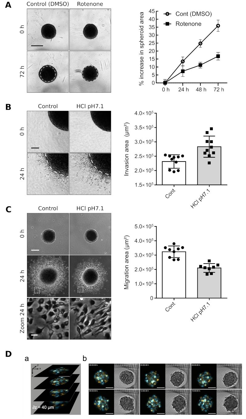
      4. Incubate the collagen gel for 30 min at 37 °C and then add cNBM on top of the gel. Inhibitors or activators (e.g., hydrogen chloride as shown in Figure 1B, 1C) can be added to the medium at this step.
    2. Image acquisition and analysis
      1. Take pictures sequentially with a video microscope in brightfield mode 24 h after collagen inclusion.
      2. Use Fiji to analyze pictures either manually or in a semiautomated manner. To do so manually, draw around the core and the total area of the spheroid with the freehand selection tool and measure the invasive area of each spheroid by subtract total area with core area. To analyze the images in a semiautomated manner, use a macro to determine the invasive area.
Access restricted. Please log in or start a trial to view this content.

Results

Loading...
$$\rightleftharpoonup{xx}$$ $$\longleftharp{xx}$$, $$\longrightharp{xx}$$,

Spheroid invasion and migration under Rotenone and HCl, diagram with graphs, microscopy results.

Figure 1: Glioblastoma P3 spheroid in proliferation, invasion, or migration assays. (A) Proliferation assay. Left panel: Representative pictures with DMSO as control or with 20 µM of rotenone (respiratory chain...

Access restricted. Please log in or start a trial to view this content.

Materials

List of materials used in this article
NameCompanyCatalog NumberComments
96 well round-bottom plateFalcon08-772-212
AccutaseGibcoA11105-01Stored at 4 °C, sphere dissociation enzyme
B27Gibco12587Stored at -20 °C, defrost before use
Basic Fibroblast Growth FactorPeprotech100-18BStored at -20 °C, defrost before use
DPBS 10XPan BiotechP04-53-500Stored at 4 °C
MethylcelluloseSigmaM0512Diluted in NBM for a 2% final concentration
NBMGibco21103-049Stored at 4 °C
Neurobasal mediumGibco21103049Stored at 4 °C
Penicillin - StreptomycinGibco15140-122Stored at 4 °C
Type I CollagenCorning354236Stored at 4 °C

Reprints and Permissions

Request permission to reuse the text or figures of this JoVE article

Request Permission

Tags

3D Spheroid ModelTumor Cell InvasionGlioblastoma ModelCollagen Matrix EmbeddingMethylcellulose SuspensionDissociation Enzyme TreatmentSpheroid Formation ProtocolExtracellular Matrix DegradationProtease Secretion AnalysisInvasive Phenotype Assessment

Related Articles