This video demonstrates the procedure to induce a closed-head injury in a mouse model using a stereotaxic frame. The controlled impact replicates mild traumatic brain injury, allowing researchers to study neurological effects and recovery by measuring post-injury reflexes and behavior.
A subscription to JoVE is required to view this content. Sign in or start your free trial.
Method Article
Induction of Mild Traumatic Brain Injury in a Mouse Model
1K views
⸱
April 28th, 2025
In This Article
Abstract
Protocol
All procedures involving animal models have been reviewed by the local institutional animal care committee and the JoVE veterinary review board.
1. Surgical procedure
- Secure the mouse into the stereotaxic frame using lightweight acetal resin tapered point ear bars, a bite bar, and a mouse anesthesia mask (see Figure 1G, H). The isoflurane gas is delivered at 2%-3% in room air at 100-200 mL/min. Carefully monitor the mouse's respiration to ensure the depth of anesthesia and adjust the level of gas as needed.
- Apply sterile eye lubricant to the eyes to prevent corneal drying.
- Sterilize the scalp with povidone-iodine swabs and sterile alcohol pads three times.
- Ensure the mouse is deeply anesthetized by verifying the lack of a toe-pinch response.
- Make an approximately 1 cm midline scalp incision between the eyes and neck using a scalpel, exposing the skull (see Figure 1I).
- Allow the skull to dry for 1-2 min.
- Identify bregma (the intersection point of the coronal and sagittal sutures) and lambda (the intersection of the sagittal and lambdoid sutures) (see Figure 1J).
NOTE: A mouse brain atlas could be used for reference. - Place the head support apparatus under the head and inflate the bulb with water until it is pressing against the bottom of the mouse's head but not lifting the head away from the bite bar.
NOTE: This step is essential to reduce possible ear issues from the closed-head injury (CHI). Any animal with damage to the ear from the ear bars, resulting in rolling or bleeding, should be eliminated from the study and euthanized. - Move the impactor into place over the animal's head.
- Extend the impactor by placing the Extend/Retract toggle switch (on the impactor control box) on the Extend.
NOTE: Be sure to check that the tip is fully extended by pulling down on the tip. - Line up the impactor until it is centered over the bregma (see Figure 1K).
- Reset the digital stereotaxic x and y coordinates in the stereotaxic reader to 0 (on the touch screen control)
- Align the probe over the impact location by moving the probe from the bregma over to the target coordinates: medial-lateral = 0.0 mm, anterior-posterior = −1.6 mm.
- Clip the contact sensor to the animal's ear.
- Slowly lower the probe tip with the extended probe until first contact with the surface is made. Stop at the beep.
- Reset the digital stereotaxic z coordinates in the stereotaxic reader to 0.
- Carefully inspect if the tip is flush with the skull (medial-lateral and anterior-posterior planes).
NOTE: Positioning the probe tip is the most crucial step of this process to prevent skull fractures and ear damage. - Retract the impactor by placing the toggle switch on the control box in the Retract position. The tip withdraws and has no more contact with the animal's head until the time of impact.
- Set the impact depth by adjusting the dorsal-ventral depth to −1.2 mm.
NOTE: The depth of the impact affects the severity of the injury. The depth should be titered for different ages, weights, and strains of mice to the desired injury severity. The depth may need to be adjusted/re-titered over time to maintain a consistent injury severity. The severity can be assessed neuropathologically: microglia and astrocytes (Immunohistochemistry (IHC)) and behaviorally: the radial arm water maze and the active avoidance test. - Carefully monitor the mouse's respiration to ensure the anesthesia depth and adjust the gas level as needed.
NOTE: Often, the percentage of isoflurane gas should be lowered or shut off for 10-20 s prior to impact. Watch closely for the respiration to accelerate slightly. If respiration is too slow at the time of impact, the animal may die within the first 60 s after the impact from apnea. This can be prevented by adjusting the depth of anesthesia in the seconds before the impact. - Induce the impact by pressing the right toggle switch to impact. The probe tip goes down at the displayed velocity and then stays down for the set dwell time and retracts.
NOTE: Sham mice receive identical handling to the CHI mice, but the impact is not delivered. - Start the timer immediately after the CHI impact is delivered to record the righting times (time to return from the side position to the prone position) or start the timer when the mouse is removed from the stereotaxic frame for the sham mice. The average righting reflex time is 5-15 min.
NOTE: The righting reflex times may vary based on mouse strain and age. - Evaluate the mice for visible skull fractures, hemorrhages, and apnea. Exclude the mice with a depressed skull fracture or visible hemorrhage from the study.
NOTE: There are graded levels of skull fractures. Animals with decompressed skull fractures, where the bone is observably pressing into the brain tissue, are euthanized (CO2 first, and decapitation is used as a secondary method). If the impactor tip is set correctly, these types of skull fractures are exceedingly rare. If a skull fracture does occur, the more common presentation is a small drop of blood on the skull and a slight tactile roughing of the skull, often along the suture connecting the posterior tip of the nasal bone. These mice are noted as possible skull fractures in the records but are not normally excluded from the study. - Remove the animal from the stereotaxic frame.
- Close the scalp by stapling the skin together.
NOTE: Absorbable or non-absorbable sutures could be used to close the scalp as an alternative to staples. - Apply triple antibiotic ointment with a sterile cotton-tipped applicator to the closed incision.
- Return the mouse to a clean holding cage for recovery. Half of the recovery cage is on a heating pad (low setting), providing the ability to move away from the heat when awake and maintaining the animal's temperature while unconscious (see Figure 1L).
NOTE: The mouse is placed on its side in the recovery cage. To prevent suffocation, place the animal in a recovery cage without bedding or on a tissue if bedding is in the cage. - Return the Extend/Retract toggle switch to the Center/Off position.
NOTE: The current will continue running if the switch is left in either the extend or retract position, causing the piston to swell. The impactor will then not be functional until the piston cools down. - Remove the impactor from its holder and gently place it on the ice pack.
NOTE: Keeping the impactor on an ice pack helps reduce the potential swelling of the impactor. - Monitor the animal until the righting reflex occurs and document the time until righting (see Figure 1M).
NOTE: The righting reflex is defined as the moment the mouse returns to a prone position. The cage needs to be left undisturbed; the mouse could right if the cage is touched, moved, or exposed to some noises. - Return the mice to their home cage when they are awake and alert. Usually, within 1 h after injury, animals are fully conscious and ambulating. Also, add some moist food at the bottom of the cage.
Access restricted. Please log in or start a trial to view this content.
Results
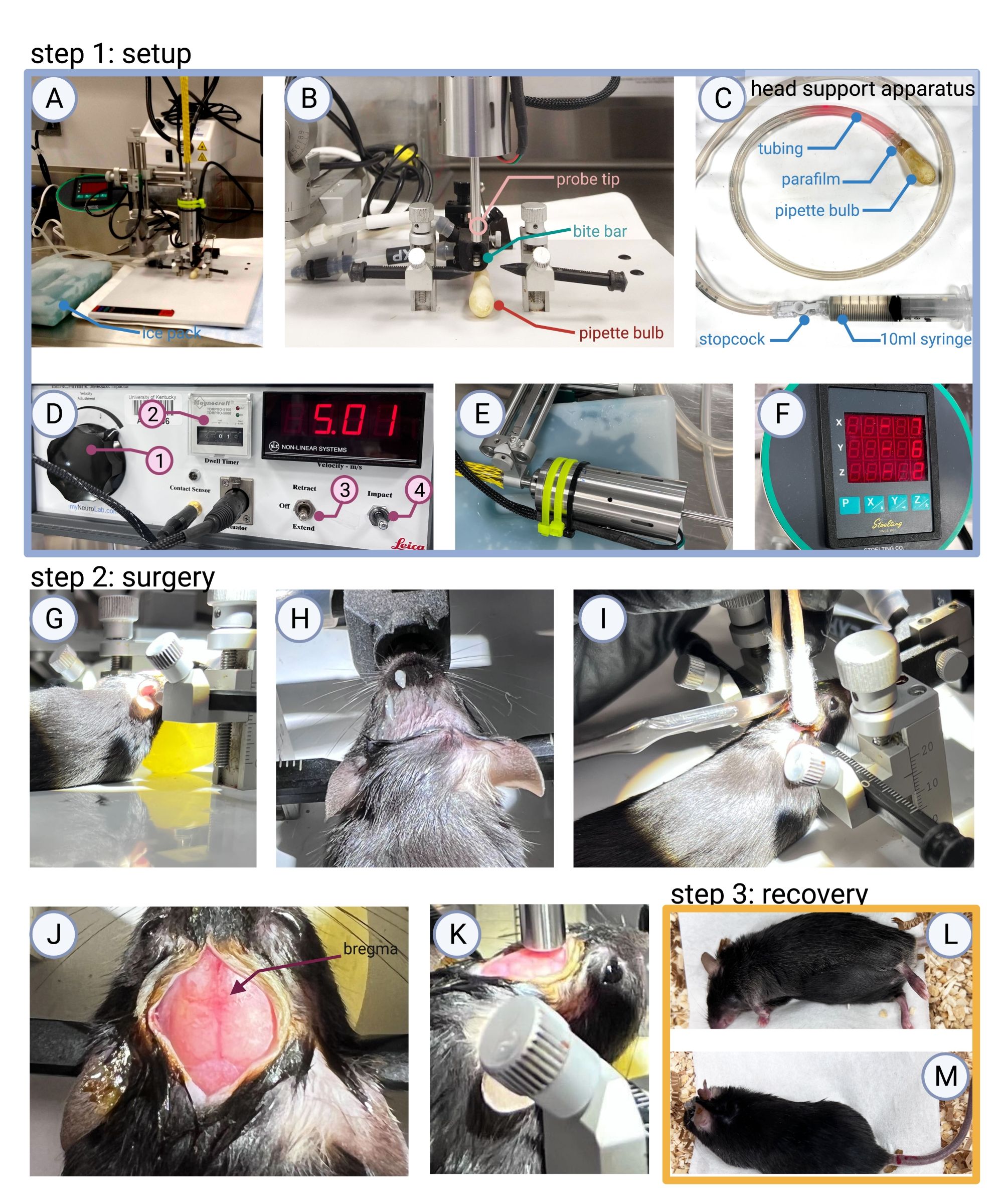
Figure 1: Step 1: Surgical area setup. (A) An example of the surgical area and tools needed to perform CHI surgery (ice pack for the impactor, stereotaxic frame equipped with the impactor, impactor control b...
Access restricted. Please log in or start a trial to view this content.
Materials
| Name | Company | Catalog Number | Comments |
|---|---|---|---|
| 9 mm Autoclip Applier | Braintree scientific | ACS- APL | Surgery |
| 9 mm Autoclip Remover | Braintree scientific | ACS- RMV | Surgery |
| 9 mm Autoclip, Case of 1,000 clips | Braintree scientific | ACS- CS | Surgery (Staples) |
| Aperio ImageScope software | Leica BioSystems | NA | IHC |
| BladeFLASK Blade Remover | Fisher Scientific | 22-444-275 | Surgery |
| Cotton tip applicator | VWR | 89031-270 | Surgery |
| Digitial mouse stereotaxic frame | Stoelting | 51730D | Surgery |
| Dumont #7 Forceps | Roboz | RS-5047 | Surgery |
| Ear bars | Stoelting | 51649 | Surgery |
| EthoVision XT 11.0 | Noldus Information Technology | NA | RAWM |
| Fiber-Lite | Dolan-Jeffer Industries | UN16103-DG | Surgery |
| Fisherbrand Bulb for Small Pipets | Fisher Scientific | 03-448-21 | Head support apparatus |
| Gemini Avoidance System | San Diego Instruments | NA | Active avoidance |
| Heating Pad | Sunbeam | 732500000U | Surgery prep |
| HRP conjugated goat anti-rabbit IgG | Jackson Immuno Research laboratories | 111-065-144 | IHC |
| Induction chamber | Kent Scientific | VetFlo-0530XS | Surgery prep |
| Isoflurane, USP | Covetrus | NDC: 11695-6777-2 | Surgery |
| Mouse gas anesthesia head holder | Stoelting | 51609M | Surgery |
| Neuropactor Stereotaxic Impactor | Neuroscience Tools | n/a | Surgery: Formally distributed by Lecia as impact one |
| NexGen Mouse 500 | Allentown | n/a | Post-surgery, holding cage |
| Parafilm | Bemis | PM992 | Head support apparatus |
| Peanut - Professional Hair Clipper | Whal | 8655-200 | Surgery prep |
| Povidone-Iodine Solution USP, 10% (w/v), 1% (w/v) available Iodine, for laboratory | Ricca | 3955-16 | Surgery |
| Puralube Vet Oinment,petrolatum ophthalmic ointment, Sterile ocular lubricant | Dechra | 17033-211-38 | Surgery |
| Rabbit anti-GFAP | Dako | Z0334 | IHC |
| Rabbit anti-IBA1 | Wako | 019-19741 | IHC |
| 8-arm Radial Arm Water Maze | MazeEngineers | n/a | RAWM |
| Scale | OHAUS CS series | BAL-101 | Surgery prep |
| Scalpel Handle #7 Solid 6.25" | Roboz | RS-9847 | Surgery |
| Sterile Alcohol Prep Pads (isopropyl alcohol 70% v/v) | Fisher Brand | 22-363-750 | Surgery prep |
| SumnoSuite low-flow anesthesia system | Kent Scientific | SS-01 | Surgery |
| 10 mL syringe Luer-Lok Tip | BD Bard-Parker | 302995 | Head support apparatus |
| Timers | Fisher Scientific | 6KED8 | Surgery |
| Topical anesthetic cream | L.M.X 4 | NDC 0496-0882-15 | Surgery prep |
| Triple antibiotic ointment | Major | NDC 0904-0734-31 | Post-surgery |
| Tubing | MasterFlex | 96410-16 | Head support apparatus |
| Vaporizer Single Channel Anesthesia System | Kent Scientific | VetFlo-1210S | Surgery prep |
