This video demonstrates the method for inducing a contusion injury at the T9 level spinal cord in a mouse model, using a spinal cord injury coaxial platform. A weight is dropped on the T9 level spinal cord that creates a contusion that immediately causes tissue discoloration and swelling.
A subscription to JoVE is required to view this content. Sign in or start your free trial.
Method Article
Inducing a Spinal Cord Contusion in a Mouse Model Using a Spinal Cord Injury Coaxial Platform
1.6K views
⸱
April 28th, 2025
In This Article
Abstract
Protocol
All procedures involving animal models have been reviewed by the local institutional animal care committee and the JoVE veterinary review board.
1. Locating and laminectomy of the 9th thoracic vertebra (T9)
NOTE: Female C57BL/6J mice 9-10 weeks old were purchased from Jinan Pengyue Experimental Animal Company (Jinan, China).
- Autoclave a suite of surgical instruments for the experiment and sterilize the operating table with 75% alcohol before surgery.
- Inject buprenorphine for analgesia (0.05-2.0 mg/kg, subcutaneously) 30 minutes before anesthesia for injury operations. Then, anesthetize the mouse with isoflurane (induction: ~3-5%, maintenance: ~1.5-2%). Check if the animal is fully anesthetized by the reflexes of the tail or toe pinch. Once anesthesia is in effect, lay the mouse in a prone position in a designated part of the operating table and coat the cornea with ophthalmic ointment (apply ophthalmic ointment to the corneas to protect the eyes from drying during surgery).
- Shave the hair from the caudal to the rostral with an electric shaver over the thoracolumbar spine. Sterilize the skin several times in a circular motion with iodophor for 30 s, followed by 75% alcohol. Apply a sterile surgical drape and make a longitudinal incision of approximately 1.5 cm on the skin from approximately the 6th thoracic vertebra (T6) to the 13th thoracic vertebra (T13) with a scalpel and blade.
NOTE: Palpate along the costal margin to the midline, where the 12th thoracic vertebra (T12) - T13 interspinous space is located. Make a 1.5 cm incision to the rostral, and the incision is approximately flush with T6-T13 vertebrae.
- Shave the hair from the caudal to the rostral with an electric shaver over the thoracolumbar spine. Sterilize the skin several times in a circular motion with iodophor for 30 s, followed by 75% alcohol. Apply a sterile surgical drape and make a longitudinal incision of approximately 1.5 cm on the skin from approximately the 6th thoracic vertebra (T6) to the 13th thoracic vertebra (T13) with a scalpel and blade.
- Explore the 13th rib on one side from the bony portion under the operating microscope. Explore the spinous process in the midline by lightly touching the area of the costovertebral angle and then toward the rostral to locate the interspinous space of the T12-T13. Explore the interspinous space of the T9 - 10th thoracic vertebra (T10) from the space of the T12-T13 to the rostral side. (Figure 1A, 2A)
- Dissect the paraspinal muscle along the spinous process of the T9 to the anterior and posterior facet joints of both sides with micro scissors (Figure 2B). Retract the paraspinal muscles with micro-retractors and clean the soft tissue on the lamina and in the interspinous space of the 8th thoracic vertebra (T8) - T9 and T9-T10 with micro scissors.
- Perform T9 laminectomy, clamp the spinous process of T9 with microsurgery forceps, slightly lift it up, insert the micro scissors parallelly along the right dorsolateral side of the lamina, avoiding damage to the spinal cord, and cut off the lamina with micro scissors. Repeat on the left side, and the spinal cord can be exposed (Figure 1B, 2C).
- Before fixing the vertebra, loosen the universal arm and slowly clamp the 9th to 10th facet joints on both sides of the vertebra with the micro mosquito forceps of the vertebral stabilizer. Tighten the screws on the micro mosquito forceps, and the vertebra is thus stabilized. Adjust the spinal cord to the horizontal plane, tighten the universal arm, and fix the vertebra (Figure 2D).
2. T9 contusion injury
- Once the T9 level spinal cord is exposed and the vertebra is fixed, aim at the spinal cord by the tip inside the sleeve under the operating microscopic (Figure 2E).
- Check if the surface of the tip is parallel to the spinal cord from the posterior and lateral aspects of the spinal cord since it is easy to observe the relationship between the spinal cord and the tip under the microscope, and the operating table can be turned easily.
- Check if the surface of the tip is parallel to the bilateral borders of the spared lamina before the tip is in contact with the spinal cord after the laminectomy since it is a natural reference plane parallel to the spinal cord.
- After locating the interspinous space of the T12-T13, lower the sleeve until the end of the impactor is consistent with the mark on the observation window and the specified height of 22 mm is reached. Pull out the pull pin to release the weight (1.3 g, 2.0 g, or 2.7 g according to the group, with each group including 3 mice and each group having one mouse for each time point).
NOTE: The spinal cord should be parallel to the ground and perpendicular to the tip; move the operating table to ensure the microscopic visual field, since the table is very compact. - Remove the impactor when the contusion is done and observe the degree of spinal cord injury (SCI) under the operating microscope. In the mild group, a light red color alteration can be seen, while in the moderate group, the injury site exhibits dark red in 3-4 s, and, possibly, eminence can be observed. In the severe group, the dark red manifestations may appear immediately, and obvious eminence in the dura is manifested, but the dura is still in a consistent shape (Figure 2F).
Access restricted. Please log in or start a trial to view this content.
Results
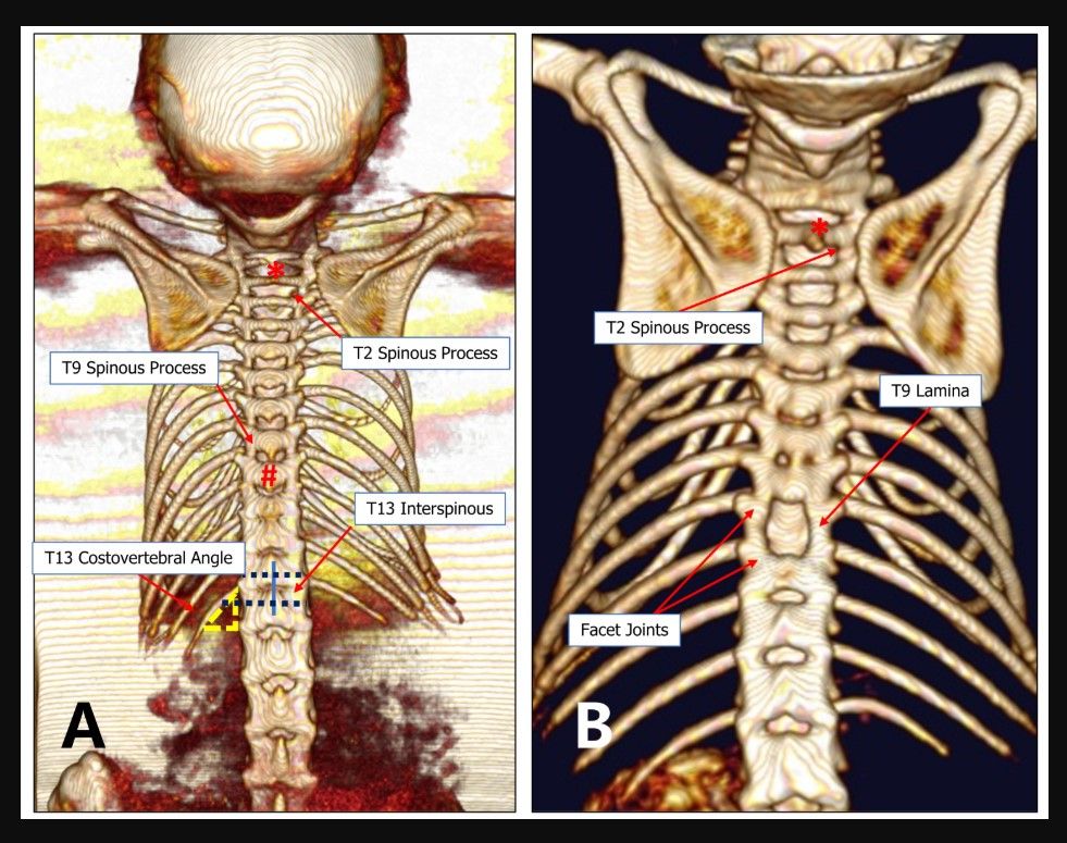
Figure 1. An imaging graph of the T13 costovertebral vertebra locating method. (A) The 13th rib and T13 are relatively constant anatomical structures. The T13 costoverte...
Access restricted. Please log in or start a trial to view this content.
Materials
| Name | Company | Catalog Number | Comments |
|---|---|---|---|
| Ethanol | Fuyu Reagent | 64-17-5 | |
| Fluorescent microscope | KEYENCE | BZ-X800 | |
| Mice | Jinan Pengyue Experimental AnimalCompany | C57BL/6J | |
| Microsurgery apparatus | Shandong ULT Biotechnology Co., Ltd | All the surgey instruments are custom-made | Ophthalmic scissors, micro mosquito forceps, microsurgery forceps, micro scissors |
| RWD Laboratory inhalation anesthetic station | RWD Life Science Co., Ltd | R550 | |
| Small animal in vivo microCT imaging system | PerkinElmer | Quantum GX2 | |
| Spinal cord injury coaxial platform | Shandong ULT Biotechnology Co., Ltd | Custom-made (Feng's standard) | (https://shop43957633.m.youzan.com/wscgoods/detail/367x5ovgn69q18g?banner_id=f.81386274~goods.7~1~ b0yRFKOq&alg_id=0&slg=tagGood List-default%2COpBottom%2Cuuid %2CabTraceId&components_style_ layout=1&reft=1659409105184&sp m=g.930111970_f.81386274&alias =367x5ovgn69q18g&from_uuid=136 2cc46-ffe0-6886-2c65-01903dbacbb a&sf=qq_sm&is_share=1&shopAuto Enter=1&share_cmpt=native_ wechat&is_silence_auth=1) |
