This video demonstrates monitoring interneuromast cell recovery in zebrafish using confocal microscopy, tracking cell death and repair through time-lapse fluorescence imaging.
A subscription to JoVE is required to view this content. Sign in or start your free trial.
Method Article
Monitoring Recovery of Laser-Ablated Interneuromast Cells in Zebrafish Using Confocal Microscopy
644 views
⸱
May 29th, 2025
In This Article
Abstract
Protocol
1. Post-ablation imaging and time-lapse microscopy to study regeneration
- Click on the dropdown menu for “Channels”. In channels, unclick the “DAPI”(4′,6-diamidino-2-phenylindole) track to inactivate the ablation laser. Click on the dropdown menu for “Acquisition mode”.
- In acquisition mode, click on “Zoom”.
- Decrease the zoom to 0.7 either by using the slider or by typing in “0.7.”
- To ensure successful cell ablation, increase the gain to 900 and fast scan the field of view.
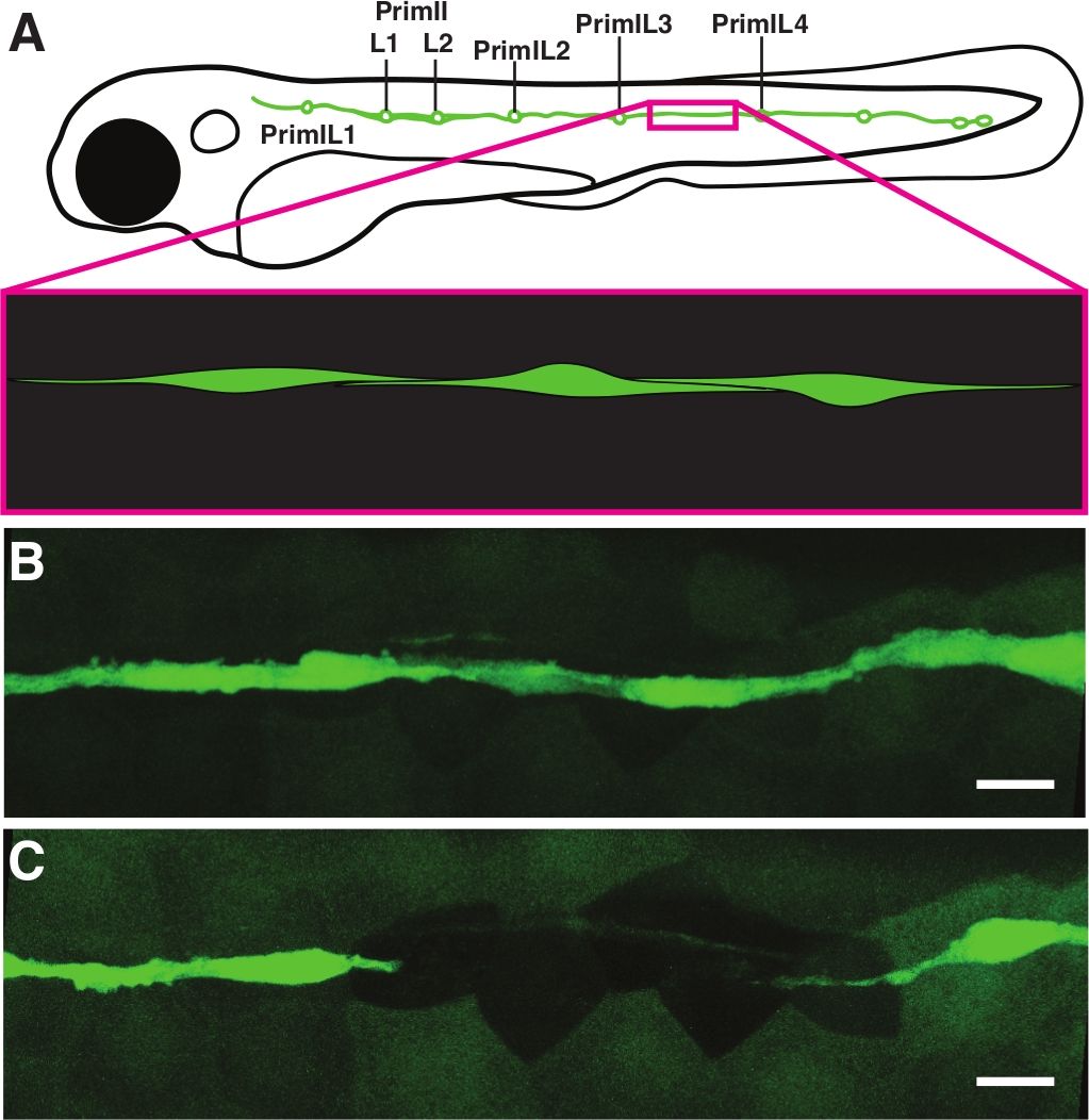
NOTE: No GFP (Green fluorescent protein)should be visible within the targeted cell or cells. Using the same or similar settings as those described for pre-ablation imaging, capture and save a post-ablation image (Figure 1C). If the image reveals remnants of fluorescent cells or additional cell bodies that needs to be removed, repeat the laser ablation steps to create a visible gap in the string of interneuromast cells. - Inspect the T-PMT (Transmission Photomultiplier Tube) channel image to confirm cellular damage further. Damaged cells will have a granular appearance, and the nuclei will frequently swell or become irregular in shape (Figure 2).
- To ensure successful cell ablation, increase the gain to 900 and fast scan the field of view.
- After capturing a pre-ablation image, ablating the cells as necessary, and capturing a post-ablation image for each individual stage position, set up the time-lapse microscopy by activating both stage position and time options for image capture. Set the time parameters to 24 h or another desired endpoint and 15 min intervals. Start experiment to acquire images and save the resulting file when complete (on the following day).
- If larvae are to be further studied or raised, carefully remove them from the agarose using fine forceps, then transfer them into E3 medium without tricaine for recovery. If they are not to be used further, euthanize according to the approved animal protocol (rapid and sustained immersion in an ice bath is a common method) and dispose of them as required by the institution.
Access restricted. Please log in or start a trial to view this content.
Results
Access restricted. Please log in or start a trial to view this content.
Materials
| Name | Company | Catalog Number | Comments |
|---|---|---|---|
| 12-well PTFE Printed Slides | |||
| 15 mM Tricaine stock solution | |||
| 1X E3 + 600 µM Tricaine | |||
| 1X E3 media | |||
| 60 X E3 media | |||
| Bx60 Compound microscope with mercury arc lamp fluorescence | |||
| LSM 700 confocal microscope equipped with 405 nm, 488 nm, and 555 or 561 nm lasers | |||
| FIJI/ImageJ Image processing software | |||
| Dissecting needle modified into hair knife | |||
| Microsoft Excel software | |||
| Glass bottom 35 mm dishes with no. 1.5 coverslip, 20 mm window | |||
| Immersol 518F Immersion Oil | |||
| Low Gelling Agarose | |||
| Corning Netwell Insert with 74 um Polyester Mesh, 24 mm Insert | |||
| Rstudio software | |||
| SMX-168-BL Stereo microscope | |||
| Transfer Pipette | |||
| ZEN software |
