This video demonstrates the procedure to embed the Drosophila brain in a fibrin matrix, followed by time-lapse fluorescence imaging of circadian clock neurons to analyze rhythmic activity synchronized with light/dark cycles.
A subscription to JoVE is required to view this content. Sign in or start your free trial.
Method Article
Embedding Drosophila Brain in a Matrix for Time-Lapse Imaging of the Circadian Clock Neurons
764 views
⸱
May 29th, 2025
In This Article
Abstract
Protocol
- Embedding of brain explants in a 3D matrix and mounting (~20 min).
- Submerge a dissected brain in fibrinogen working solution (fibrinogen final concentration ~3.3 mg/mL, 10.5 µL per brain) as follows:
- Aspirate 3.5 µL of schneider active medium (SAM)/antibiotics. Aspirate a dissected brain from the dissection well into the same pipette tip without dispensing the SAM/antibiotics medium in the tip.
- Dispense the brain and the medium on the lid of a sterile Petri dish. Add another 3.5 µL of SAM/antibiotics and 3.5 µL of warm fibrinogen/SAM 10 mg/mL solution to the drop containing the brain. Gently mix the solution around the brain by pipetting with a 20 µL pipette tip.
NOTE: Using a pipette rather than forceps in this step avoids stressing the brain. - Take 2 µL of the fibrinogen working solution from the drop and apply it as a small circle on the coverslip of the glass-bottom dish. Add 0.8 µL of thrombin and mix very quickly with a pipette tip. The fibrinogen is converted to fibrin and starts to polymerize.
- Take the brain that sits in the fibrinogen working solution (step 1.1) between a pair of forceps and position it on the matrix once a white matrix of fibrin is visible. Orient the brain anterior side down (for imaging clock neurons). Push it down as close as possible to the cover glass. The fibrin clot is composed of layers of fibrin. Pick one layer and fold it on top of the brain with small and gentle movements without detaching the matrix.
- Repeat 2 to 3 times from different parts of the clot to stabilize the brain.
NOTE: When handling the brain with forceps, be careful not to dry it or impose physical stress on it.
- Repeat 2 to 3 times from different parts of the clot to stabilize the brain.
- Add 20 µL of SAM/antibiotics on top of the embedded brain to stop the action of thrombin.
- Repeat the procedure to mount the remaining brains. Up to five to six brains can be embedded in a glass-bottom dish.
- For long-term live imaging, add 600 µL of SAM/antibiotics in the inner well (the glass part). Position a pre-cut Polytetrafluoroethylene (PTFE) membrane on top of the medium and stick it to the vacuum grease applied on the plastic part of the bottom (Figure 1A).
Access restricted. Please log in or start a trial to view this content.
Results
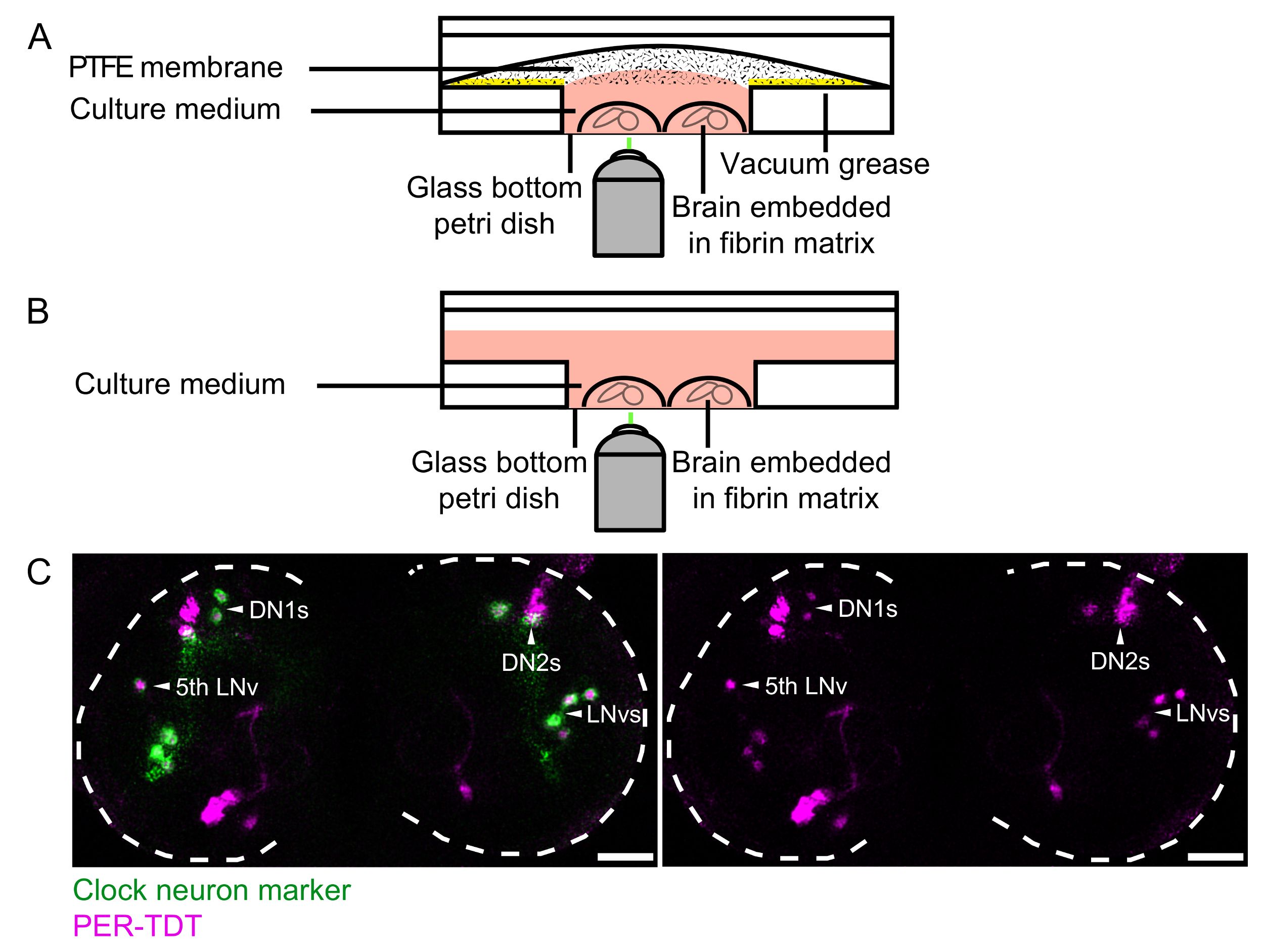
Access restricted. Please log in or start a trial to view this content.
Materials
| Name | Company | Catalog Number | Comments |
|---|---|---|---|
| KH2PO4 | |||
| CaCl2 | |||
| MgSO4.7H2O | |||
| NaCl | |||
| NaHCO3 | |||
| D-(+) Glucose | |||
| Yeast extract | |||
| Insulin | |||
| Penicillin-Streptomycin | |||
| BIS-TRIS | |||
| L-(−)-Malic acid | |||
| D-(+)-Trehalose dihydrate | |||
| Succinic acid | |||
| Fumaric acid | |||
| α-Ketoglutaric acid | |||
| Non-heat-inactivated, Foetal Calf Serum (FCS) Mycoplasma and Virus screened | |||
| HEPES-KOH, pH 7.4 | |||
| KCl | |||
| NaH2PO4 | |||
| Sucrose | |||
| Fibrinogen from bovine plasma | |||
| Thrombin from bovine plasma | |||
| PDF, NH2-NSELINSLLSLPKNMNDA-OH | |||
| Vaccum grease | |||
| 35 mm Dish, No. 1.5 Coverslip, 20 mm Glass Diameter, Uncoated | |||
| Corning Falcon Easy-Grip Tissue Culture Dishes | |||
| Sterile 500 mL Steritop-GP 33 mm threaded bottle top filter, 0.22 μm | |||
| Polytetrafluoroethylene (PTFE) film | |||
| Millex-HV Syringe Filter Unit, 0.45 µm, PVDF, 33 mm, gamma sterilized | |||
| Millex-GV Syringe Filter Unit, 0.22 µm, PVDF, 33 mm, gamma sterilized | |||
| Three-well glass dissection dish | |||
| Fine forceps, size 5, Dumont | |||
| Tandem scanner inverted TCS SP5 confocal microscope, with resonant scanner and HyD photo-multiplier detectors | |||
| Temperature control chamber | |||
| Stage-top humidity controller | |||
| Water Immersion Micro Dispenser: dispenser, extended micro-pump MP6 series and Autoimmersion Objective Controller software | |||
| SUM-stack creation and 3D correction drift plugin | |||
| 10x iterative deconvolution |
